Science
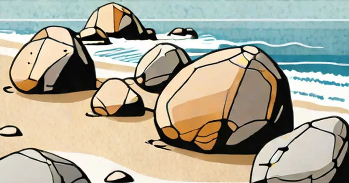
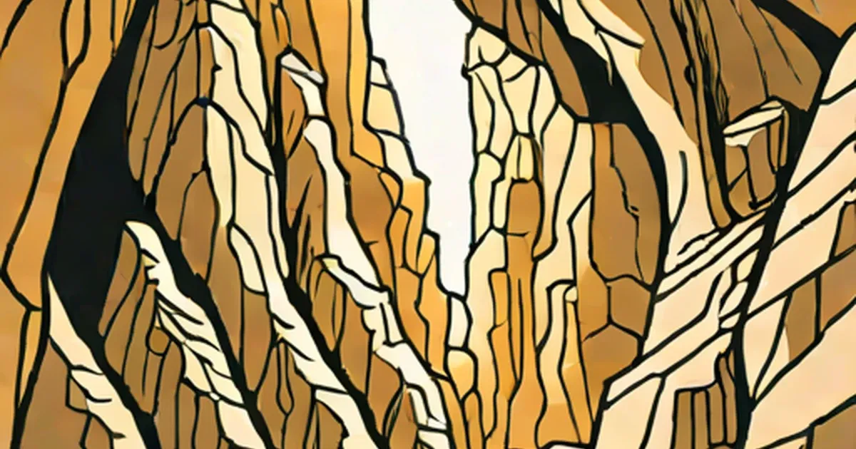
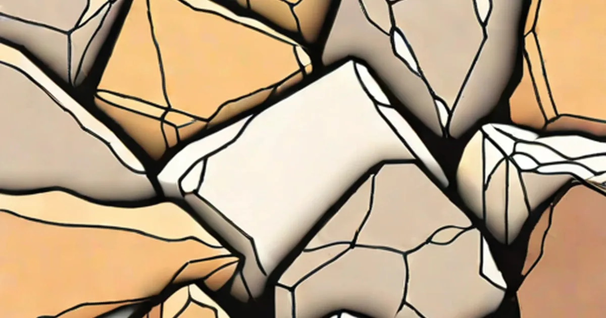
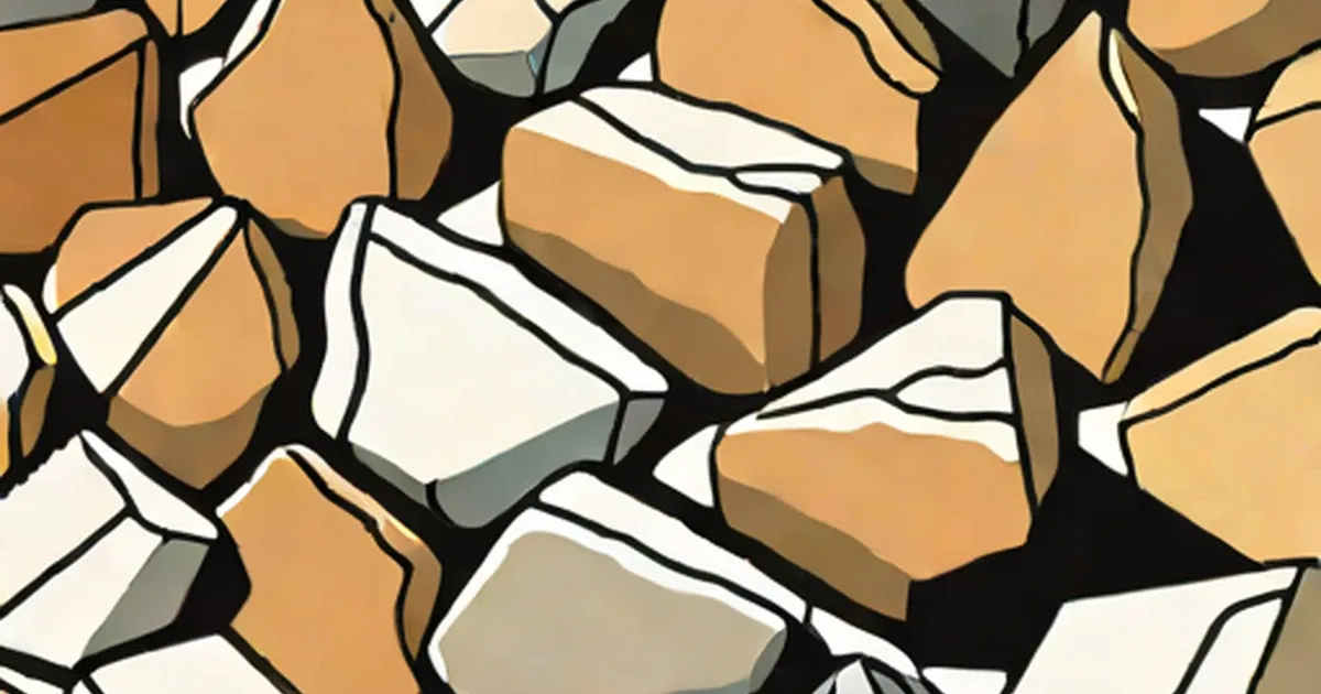
Interesting Facts About Moeraki Boulders
April 15, 2016
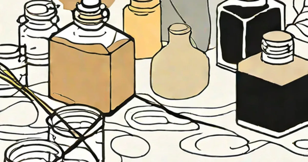
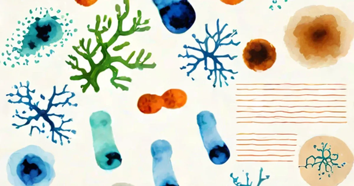
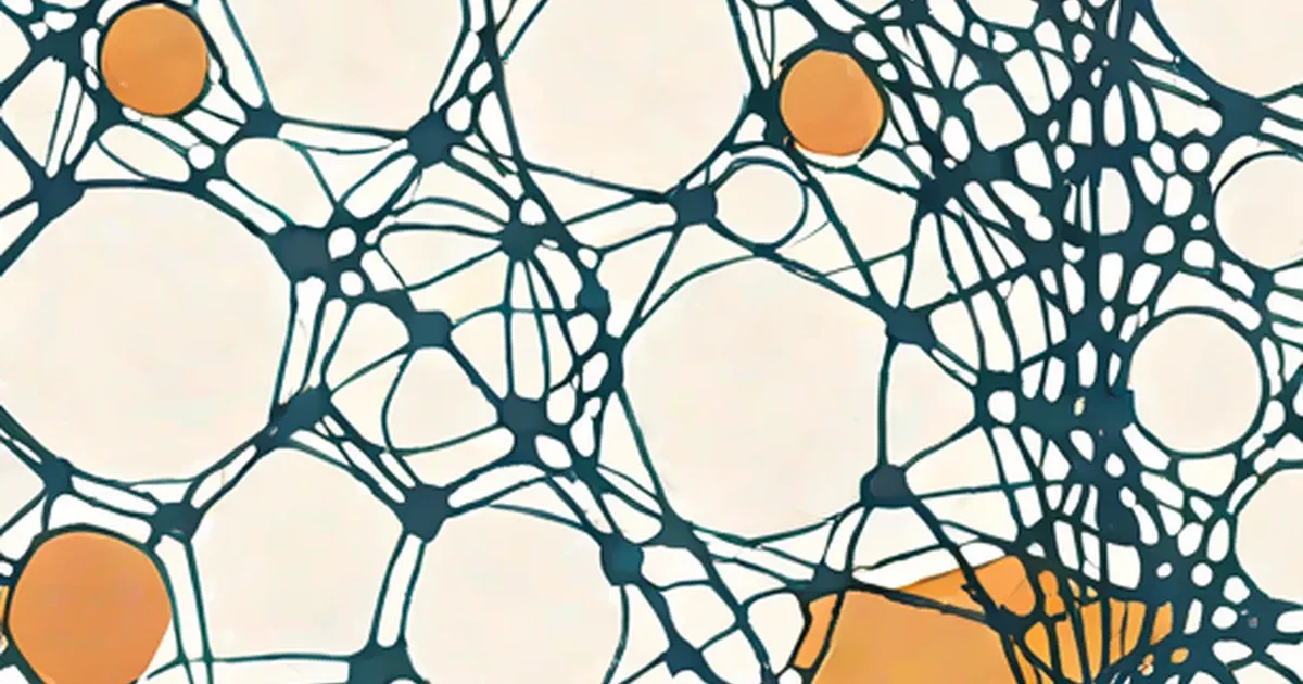
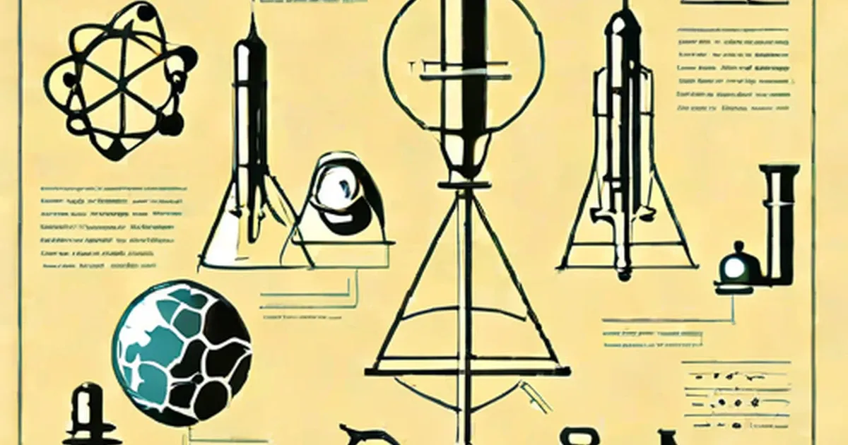
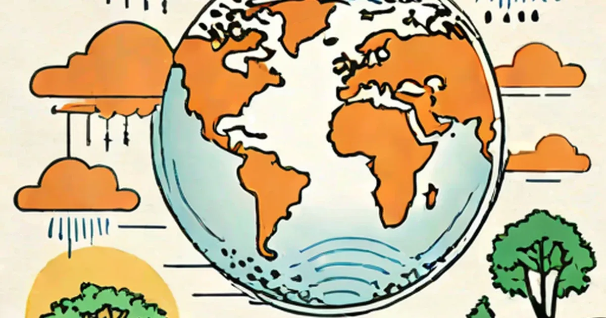
Science made clear — space, biology, chemistry, physics and earth science, explained so the ideas land without a degree in the subject.
Science
April 15, 2016
Science
April 15, 2016
Science
April 14, 2016

Science
April 14, 2016
Science
October 17, 2015
Science
October 13, 2015
Science
October 5, 2015

Science
September 19, 2015
Science
September 19, 2015
Science
September 19, 2015
Science
September 19, 2015
Science
September 18, 2015
Science
September 10, 2015
Science
September 9, 2015

Science
September 8, 2015
Science
May 23, 2015
Science
May 23, 2015
Science
August 26, 2014
Science
August 26, 2014
Science
July 13, 2014
Science
July 13, 2014
Science
June 1, 2014
Science
March 24, 2014
Science
June 13, 2013
Science
October 7, 2012
Science
May 31, 2012
Science
April 4, 2012
Science
November 23, 2011
Science
November 13, 2011
Science
November 5, 2011
Science
October 11, 2011
Science
October 7, 2011
Science
October 6, 2011
Science
October 4, 2011
Science
September 25, 2011
Science
September 24, 2011

Science
August 9, 2011
Science
August 7, 2011
Science
June 12, 2011
Science
March 25, 2011
Science
February 13, 2011
Science
February 8, 2011

Science
January 17, 2011
Science
January 16, 2011
Science
December 11, 2010
Science
December 6, 2010
Science
December 5, 2010
Science
December 3, 2010
Science
November 25, 2010

Science
November 23, 2010
Science
November 19, 2010
Science
November 19, 2010
Science
November 14, 2010
Science
November 3, 2010
Science
September 10, 2010

Science
September 10, 2010
Science
July 20, 2010
Science
July 20, 2010
Science
July 17, 2010
Science
July 11, 2010
Science
July 3, 2010
Science
July 3, 2010
Science
June 25, 2010
Science
June 13, 2010
Science
June 6, 2010

Science
June 3, 2010
Science
May 28, 2010
Science
May 26, 2010
Science
May 16, 2010
Science
May 14, 2010
Science
May 10, 2010
Science
May 10, 2010

Science
May 10, 2010
Science
May 9, 2010

Science
May 4, 2010
Science
April 19, 2010
Science
April 16, 2010
Science
April 13, 2010
Science
April 6, 2010
Science
March 30, 2010
Science
March 30, 2010
Science
March 23, 2010
Science
March 23, 2010

Science
March 22, 2010

Science
March 6, 2010
Science
March 3, 2010
Science
February 22, 2010
Science
February 17, 2010
Science
February 12, 2010
Science
February 6, 2010
Science
February 4, 2010
Science
January 27, 2010

Science
January 24, 2010
Science
January 24, 2010
Science
January 24, 2010

Science
January 22, 2010

Science
January 21, 2010

Science
January 16, 2010
Science
January 16, 2010

Science
January 14, 2010
Science
December 19, 2009
Science
December 16, 2009
Science
December 13, 2009
Science
December 8, 2009
Science
December 8, 2009
Science
December 7, 2009

Science
November 26, 2009
Science
November 22, 2009
Science
November 20, 2009
Science
November 14, 2009
Science
November 7, 2009
Science
November 7, 2009

Science
November 7, 2009
Science
November 2, 2009
Science
October 16, 2009
Science
October 13, 2009
Science
October 11, 2009
Science
October 4, 2009

Science
September 26, 2009
Science
September 25, 2009
Science
September 18, 2009
Science
September 17, 2009
Science
August 30, 2009
Science
August 21, 2009
Science
August 14, 2009
Science
August 13, 2009
Science
August 9, 2009
Science
August 4, 2009
Science
August 1, 2009

Science
July 10, 2009

Science
July 7, 2009
Science
July 7, 2009
Science
July 6, 2009
Science
June 26, 2009

Science
June 20, 2009
Science
June 15, 2009

Science
June 14, 2009
Science
May 24, 2009
Science
May 22, 2009
Science
May 19, 2009

Science
May 19, 2009

Science
May 15, 2009
Science
May 11, 2009
Science
May 9, 2009
Science
May 4, 2009
Science
May 4, 2009
Science
May 3, 2009
Science
May 2, 2009
Science
April 29, 2009
Science
April 22, 2009

Science
April 20, 2009

Science
April 19, 2009
Science
April 6, 2009
Science
April 2, 2009
Science
March 22, 2009
Science
March 15, 2009
Science
February 20, 2009
Science
February 10, 2009
Science
February 10, 2009
Science
February 10, 2009

Science
January 30, 2009
Science
January 25, 2009
Science
January 25, 2009

Science
January 24, 2009

Science
December 18, 2008
Science
December 8, 2008
Science
December 6, 2008

Science
October 2, 2008
Science
September 17, 2008
Science
August 29, 2008

Science
August 29, 2008

Science
August 17, 2008
Science
July 8, 2008
Science
July 4, 2008
Science
May 17, 2008
Science
May 6, 2008
Science
April 25, 2008
Science
April 6, 2008
Science
April 1, 2008
Science
March 27, 2008
Science
March 25, 2008
Science
March 19, 2008
Science
March 10, 2008
Science
March 6, 2008
Science
January 23, 2008
Science
January 22, 2008

Science
January 20, 2008

Science
January 9, 2008
Science
January 8, 2008
Science
January 6, 2008
Science
December 18, 2007
Science
December 13, 2007

Science
November 29, 2007
Science
November 26, 2007
Science
November 12, 2007
Science
November 2, 2007
Science
November 1, 2007
Science
October 28, 2007
Science
October 24, 2007

Science
October 18, 2007

Science
October 12, 2007
Science
October 11, 2007
Science
August 29, 2007
Science
August 17, 2007
Science
August 12, 2007
Science
July 13, 2007
Science
June 26, 2007

Science
June 6, 2007

Science
April 27, 2007
Science
April 22, 2007
Science
November 11, 2006

Science
July 3, 2006
Science
May 13, 2006
Science
March 8, 2005
Science
February 7, 2005
Science
November 28, 2004
Science
October 31, 2004
Science
October 20, 2004

Science
September 24, 2004

Science
September 19, 2004

Science
September 18, 2004
Science
June 18, 2004
Science
January 7, 2004
Science
October 1, 2003

Science
August 1, 2003

Science
April 24, 2003

Science
February 2, 2003