
Health & Fitness
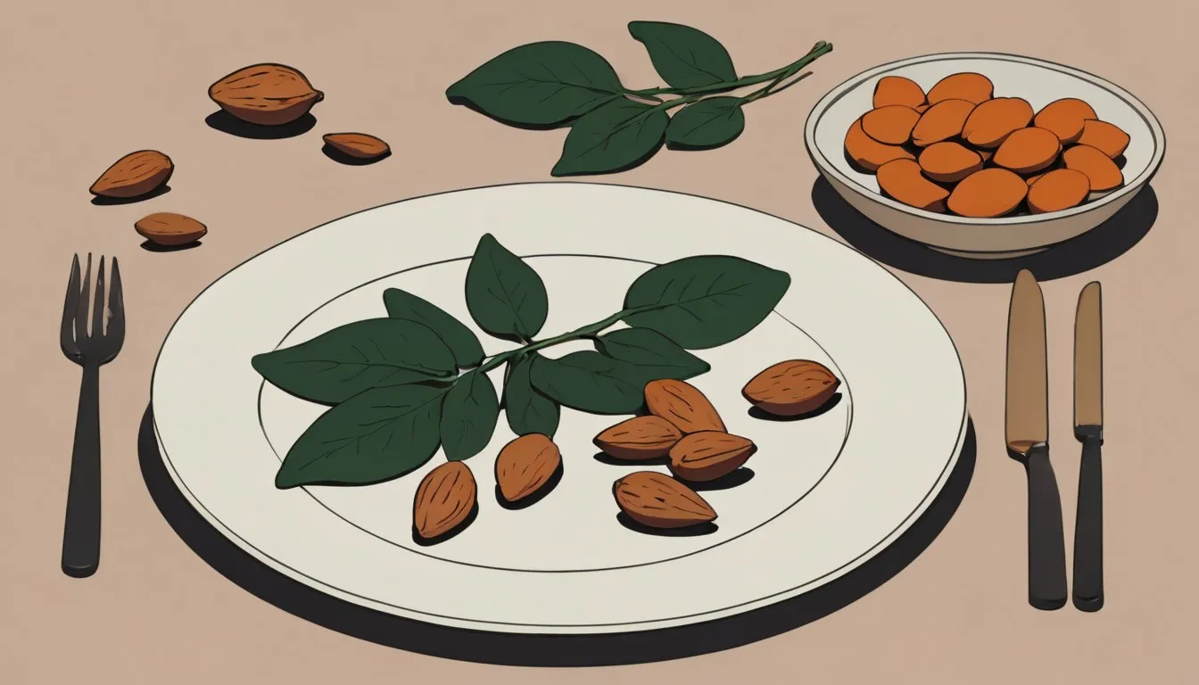
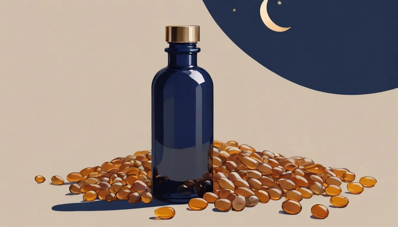
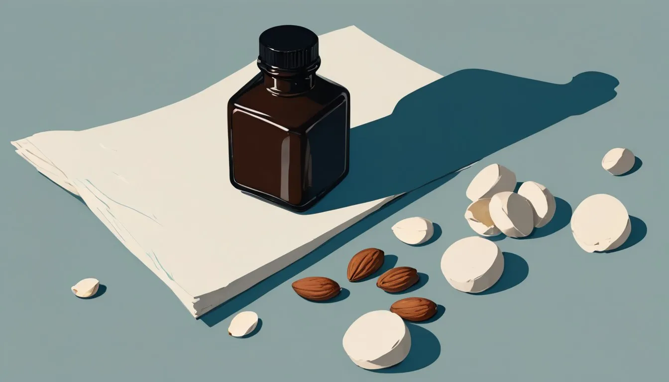
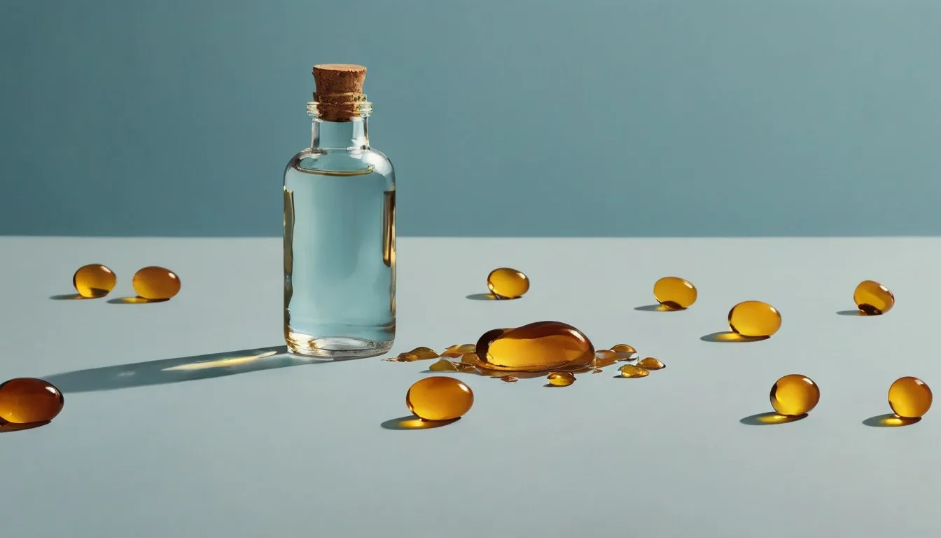
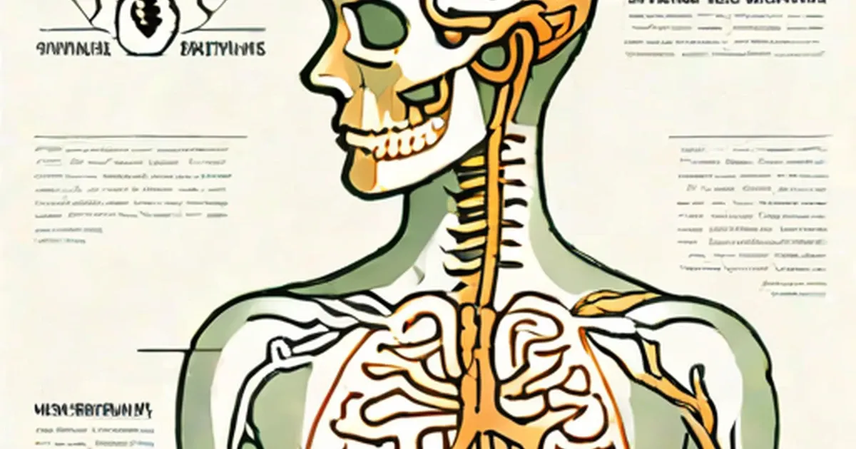
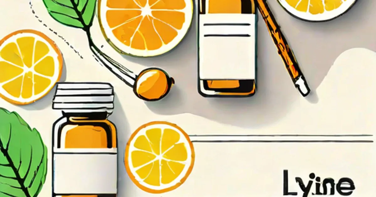
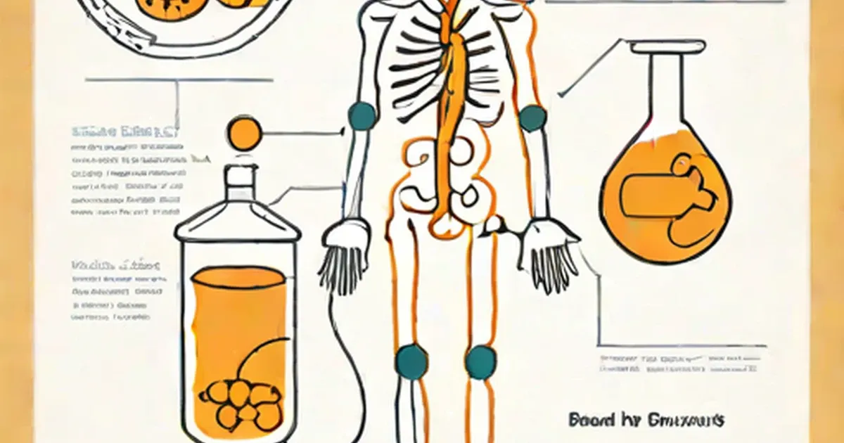
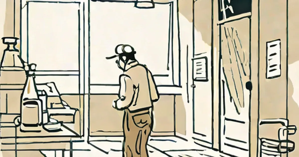
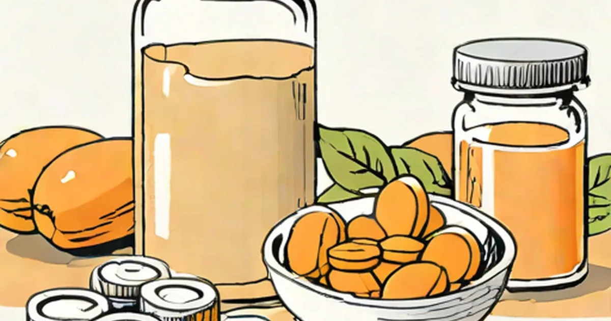
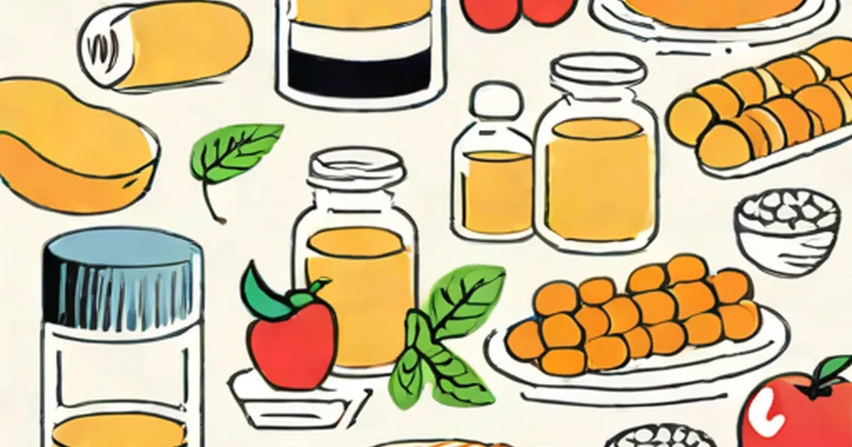
Is It Safe to Take Magnesium Supplements While Pregnant?
July 21, 2026
Health and fitness you can act on — nutrition, exercise, conditions and wellbeing, explained clearly and grounded in what the evidence supports.
Health & Fitness
July 21, 2026

Health & Fitness
July 21, 2026
Health & Fitness
July 21, 2026

Health & Fitness
July 21, 2026
Health & Fitness
July 21, 2026

Health & Fitness
July 21, 2026

Health & Fitness
July 21, 2026
Health & Fitness
July 21, 2026
Health & Fitness
July 21, 2026
Health & Fitness
July 21, 2026

Health & Fitness
July 21, 2026
Health & Fitness
July 21, 2026

Health & Fitness
July 21, 2026

Health & Fitness
July 21, 2026
Health & Fitness
July 21, 2026

Health & Fitness
July 21, 2026
Health & Fitness
July 20, 2026
Health & Fitness
July 20, 2026
Health & Fitness
July 20, 2026
Health & Fitness
July 20, 2026

Health & Fitness
July 20, 2026

Health & Fitness
July 20, 2026

Health & Fitness
July 20, 2026

Health & Fitness
July 20, 2026
Health & Fitness
July 20, 2026
Health & Fitness
July 20, 2026
Health & Fitness
July 18, 2026
Health & Fitness
July 18, 2026
Health & Fitness
July 18, 2026
Health & Fitness
July 18, 2026

Health & Fitness
July 18, 2026

Health & Fitness
July 18, 2026
Health & Fitness
July 18, 2026
Health & Fitness
July 18, 2026
Health & Fitness
July 18, 2026
Health & Fitness
July 18, 2026
Health & Fitness
July 18, 2026

Health & Fitness
July 18, 2026
Health & Fitness
July 18, 2026
Health & Fitness
July 18, 2026
Health & Fitness
July 18, 2026

Health & Fitness
July 17, 2026
Health & Fitness
July 17, 2026
Health & Fitness
July 17, 2026
Health & Fitness
July 17, 2026

Health & Fitness
July 17, 2026

Health & Fitness
July 17, 2026
Health & Fitness
July 17, 2026

Health & Fitness
July 17, 2026

Health & Fitness
July 17, 2026

Health & Fitness
July 17, 2026

Health & Fitness
July 17, 2026

Health & Fitness
July 17, 2026

Health & Fitness
July 17, 2026
Health & Fitness
July 16, 2026

Health & Fitness
July 12, 2026
Health & Fitness
July 12, 2026
Health & Fitness
July 12, 2026

Health & Fitness
April 15, 2016

Health & Fitness
April 15, 2016

Health & Fitness
April 13, 2016
Health & Fitness
November 29, 2015

Health & Fitness
November 29, 2015
Health & Fitness
November 29, 2015

Health & Fitness
October 5, 2015

Health & Fitness
October 5, 2015
Health & Fitness
October 4, 2015

Health & Fitness
September 19, 2015

Health & Fitness
September 14, 2015
Health & Fitness
April 14, 2015
Health & Fitness
September 23, 2014
Health & Fitness
May 25, 2014
Health & Fitness
August 22, 2013

Health & Fitness
June 13, 2013

Health & Fitness
March 5, 2012
Health & Fitness
November 22, 2011
Health & Fitness
November 17, 2011

Health & Fitness
October 24, 2011
Health & Fitness
October 21, 2011
Health & Fitness
October 21, 2011
Health & Fitness
October 17, 2011

Health & Fitness
October 17, 2011
Health & Fitness
October 12, 2011
Health & Fitness
October 6, 2011
Health & Fitness
October 2, 2011

Health & Fitness
September 24, 2011
Health & Fitness
August 10, 2011

Health & Fitness
August 9, 2011

Health & Fitness
July 18, 2011

Health & Fitness
July 15, 2011

Health & Fitness
June 27, 2011
Health & Fitness
June 13, 2011

Health & Fitness
June 11, 2011

Health & Fitness
June 5, 2011
Health & Fitness
June 3, 2011
Health & Fitness
May 22, 2011
Health & Fitness
May 16, 2011
Health & Fitness
May 11, 2011

Health & Fitness
May 6, 2011

Health & Fitness
May 4, 2011
Health & Fitness
May 2, 2011

Health & Fitness
April 26, 2011

Health & Fitness
April 19, 2011

Health & Fitness
April 19, 2011
Health & Fitness
April 17, 2011

Health & Fitness
April 14, 2011
Health & Fitness
April 1, 2011
Health & Fitness
March 27, 2011
Health & Fitness
March 3, 2011

Health & Fitness
February 27, 2011
Health & Fitness
February 21, 2011
Health & Fitness
February 5, 2011
Health & Fitness
January 30, 2011

Health & Fitness
January 24, 2011
Health & Fitness
January 8, 2011

Health & Fitness
December 30, 2010

Health & Fitness
December 25, 2010

Health & Fitness
December 15, 2010
Health & Fitness
December 13, 2010

Health & Fitness
December 12, 2010

Health & Fitness
December 5, 2010

Health & Fitness
November 29, 2010

Health & Fitness
November 28, 2010
Health & Fitness
November 26, 2010

Health & Fitness
November 25, 2010

Health & Fitness
November 25, 2010

Health & Fitness
November 23, 2010
Health & Fitness
November 17, 2010

Health & Fitness
November 17, 2010

Health & Fitness
November 3, 2010

Health & Fitness
October 30, 2010

Health & Fitness
October 23, 2010

Health & Fitness
October 5, 2010
Health & Fitness
October 4, 2010

Health & Fitness
September 25, 2010

Health & Fitness
September 25, 2010

Health & Fitness
September 24, 2010

Health & Fitness
September 22, 2010

Health & Fitness
September 20, 2010
Health & Fitness
September 12, 2010

Health & Fitness
September 10, 2010

Health & Fitness
August 31, 2010
Health & Fitness
August 29, 2010
Health & Fitness
August 26, 2010
Health & Fitness
August 22, 2010
Health & Fitness
August 20, 2010

Health & Fitness
August 20, 2010

Health & Fitness
August 9, 2010
Health & Fitness
July 24, 2010

Health & Fitness
July 23, 2010

Health & Fitness
July 22, 2010

Health & Fitness
July 18, 2010

Health & Fitness
July 18, 2010

Health & Fitness
July 16, 2010
Health & Fitness
July 15, 2010

Health & Fitness
July 11, 2010
Health & Fitness
July 10, 2010

Health & Fitness
July 9, 2010

Health & Fitness
July 9, 2010
Health & Fitness
July 9, 2010

Health & Fitness
July 6, 2010

Health & Fitness
June 17, 2010

Health & Fitness
June 16, 2010
Health & Fitness
June 15, 2010

Health & Fitness
June 8, 2010

Health & Fitness
June 8, 2010

Health & Fitness
June 4, 2010

Health & Fitness
June 4, 2010
Health & Fitness
May 28, 2010

Health & Fitness
May 27, 2010

Health & Fitness
May 26, 2010

Health & Fitness
May 26, 2010

Health & Fitness
May 19, 2010

Health & Fitness
May 14, 2010
Health & Fitness
May 11, 2010

Health & Fitness
May 11, 2010

Health & Fitness
May 2, 2010
Health & Fitness
April 30, 2010

Health & Fitness
April 28, 2010

Health & Fitness
April 23, 2010

Health & Fitness
April 21, 2010

Health & Fitness
April 17, 2010

Health & Fitness
April 13, 2010
Health & Fitness
April 7, 2010

Health & Fitness
April 3, 2010
Health & Fitness
April 1, 2010
Health & Fitness
March 27, 2010

Health & Fitness
March 14, 2010

Health & Fitness
March 13, 2010

Health & Fitness
March 12, 2010
Health & Fitness
March 7, 2010
Health & Fitness
March 6, 2010

Health & Fitness
March 4, 2010

Health & Fitness
February 28, 2010
Health & Fitness
February 27, 2010

Health & Fitness
February 27, 2010

Health & Fitness
February 26, 2010

Health & Fitness
February 24, 2010
Health & Fitness
February 20, 2010
Health & Fitness
February 20, 2010
Health & Fitness
February 18, 2010
Health & Fitness
February 13, 2010

Health & Fitness
February 7, 2010

Health & Fitness
January 30, 2010
Health & Fitness
January 29, 2010
Health & Fitness
January 29, 2010

Health & Fitness
January 28, 2010

Health & Fitness
January 25, 2010
Health & Fitness
January 24, 2010

Health & Fitness
January 20, 2010
Health & Fitness
January 19, 2010

Health & Fitness
January 19, 2010

Health & Fitness
January 16, 2010

Health & Fitness
January 13, 2010
Health & Fitness
January 11, 2010

Health & Fitness
January 8, 2010

Health & Fitness
January 7, 2010

Health & Fitness
December 30, 2009
Health & Fitness
December 27, 2009

Health & Fitness
December 27, 2009

Health & Fitness
December 23, 2009

Health & Fitness
December 23, 2009
Health & Fitness
December 22, 2009

Health & Fitness
December 21, 2009

Health & Fitness
December 16, 2009

Health & Fitness
December 16, 2009
Health & Fitness
December 13, 2009

Health & Fitness
December 8, 2009
Health & Fitness
December 1, 2009

Health & Fitness
November 30, 2009

Health & Fitness
November 29, 2009

Health & Fitness
November 29, 2009

Health & Fitness
November 26, 2009
Health & Fitness
November 26, 2009

Health & Fitness
November 21, 2009

Health & Fitness
November 15, 2009

Health & Fitness
November 14, 2009

Health & Fitness
November 13, 2009

Health & Fitness
November 12, 2009

Health & Fitness
November 10, 2009
Health & Fitness
November 7, 2009
Health & Fitness
November 5, 2009
Health & Fitness
October 31, 2009

Health & Fitness
October 27, 2009
Health & Fitness
October 27, 2009

Health & Fitness
October 26, 2009
Health & Fitness
October 25, 2009
Health & Fitness
October 20, 2009
Health & Fitness
October 19, 2009
Health & Fitness
October 16, 2009

Health & Fitness
October 12, 2009

Health & Fitness
October 8, 2009

Health & Fitness
October 8, 2009

Health & Fitness
October 3, 2009
Health & Fitness
September 26, 2009

Health & Fitness
September 25, 2009

Health & Fitness
September 24, 2009

Health & Fitness
September 24, 2009

Health & Fitness
September 24, 2009
Health & Fitness
September 24, 2009
Health & Fitness
September 17, 2009

Health & Fitness
September 14, 2009

Health & Fitness
September 13, 2009

Health & Fitness
September 3, 2009

Health & Fitness
August 27, 2009

Health & Fitness
August 18, 2009

Health & Fitness
August 16, 2009
Health & Fitness
August 14, 2009

Health & Fitness
August 14, 2009

Health & Fitness
August 14, 2009
Health & Fitness
July 29, 2009

Health & Fitness
July 28, 2009
Health & Fitness
July 26, 2009

Health & Fitness
July 22, 2009
Health & Fitness
July 21, 2009
Health & Fitness
July 19, 2009
Health & Fitness
July 18, 2009

Health & Fitness
July 13, 2009
Health & Fitness
July 10, 2009

Health & Fitness
July 10, 2009

Health & Fitness
July 9, 2009
Health & Fitness
July 7, 2009

Health & Fitness
July 6, 2009

Health & Fitness
June 29, 2009
Health & Fitness
June 28, 2009
Health & Fitness
June 28, 2009
Health & Fitness
June 28, 2009

Health & Fitness
June 27, 2009
Health & Fitness
June 25, 2009

Health & Fitness
June 25, 2009
Health & Fitness
June 24, 2009

Health & Fitness
June 18, 2009

Health & Fitness
June 17, 2009
Health & Fitness
June 16, 2009

Health & Fitness
June 15, 2009

Health & Fitness
June 14, 2009
Health & Fitness
June 8, 2009

Health & Fitness
June 5, 2009

Health & Fitness
May 5, 2009
Health & Fitness
May 4, 2009
Health & Fitness
May 4, 2009

Health & Fitness
April 28, 2009
Health & Fitness
April 21, 2009

Health & Fitness
April 19, 2009

Health & Fitness
April 16, 2009

Health & Fitness
March 30, 2009

Health & Fitness
March 28, 2009

Health & Fitness
March 24, 2009

Health & Fitness
March 21, 2009
Health & Fitness
March 21, 2009

Health & Fitness
March 17, 2009

Health & Fitness
March 12, 2009
Health & Fitness
February 12, 2009

Health & Fitness
January 25, 2009
Health & Fitness
December 12, 2008
Health & Fitness
October 7, 2008
Health & Fitness
October 6, 2008

Health & Fitness
September 17, 2008
Health & Fitness
September 14, 2008

Health & Fitness
August 26, 2008

Health & Fitness
August 17, 2008

Health & Fitness
August 3, 2008

Health & Fitness
July 23, 2008

Health & Fitness
June 24, 2008
Health & Fitness
June 19, 2008
Health & Fitness
May 28, 2008
Health & Fitness
May 21, 2008
Health & Fitness
May 6, 2008

Health & Fitness
April 20, 2008

Health & Fitness
March 28, 2008
Health & Fitness
March 27, 2008

Health & Fitness
March 27, 2008

Health & Fitness
March 21, 2008

Health & Fitness
March 19, 2008

Health & Fitness
March 10, 2008
Health & Fitness
March 2, 2008

Health & Fitness
February 11, 2008
Health & Fitness
February 1, 2008

Health & Fitness
January 17, 2008

Health & Fitness
January 9, 2008

Health & Fitness
January 8, 2008

Health & Fitness
January 4, 2008
Health & Fitness
January 2, 2008

Health & Fitness
December 31, 2007
Health & Fitness
December 31, 2007
Health & Fitness
December 21, 2007

Health & Fitness
November 20, 2007
Health & Fitness
November 20, 2007

Health & Fitness
November 7, 2007

Health & Fitness
November 2, 2007

Health & Fitness
October 28, 2007

Health & Fitness
October 28, 2007

Health & Fitness
October 28, 2007

Health & Fitness
October 19, 2007

Health & Fitness
October 19, 2007

Health & Fitness
October 18, 2007

Health & Fitness
October 18, 2007

Health & Fitness
October 17, 2007
Health & Fitness
October 16, 2007

Health & Fitness
October 16, 2007
Health & Fitness
October 16, 2007

Health & Fitness
October 14, 2007

Health & Fitness
October 12, 2007

Health & Fitness
October 12, 2007
Health & Fitness
October 12, 2007

Health & Fitness
October 12, 2007
Health & Fitness
October 12, 2007

Health & Fitness
August 23, 2007

Health & Fitness
June 11, 2007

Health & Fitness
May 23, 2007

Health & Fitness
May 8, 2007

Health & Fitness
April 27, 2007
Health & Fitness
April 27, 2007

Health & Fitness
April 21, 2007

Health & Fitness
April 20, 2007
Health & Fitness
April 16, 2007
Health & Fitness
March 27, 2007

Health & Fitness
March 19, 2007

Health & Fitness
February 28, 2007

Health & Fitness
February 2, 2007

Health & Fitness
January 4, 2007
Health & Fitness
January 4, 2007

Health & Fitness
January 3, 2007
Health & Fitness
November 14, 2006

Health & Fitness
October 12, 2006

Health & Fitness
October 8, 2006

Health & Fitness
August 31, 2006
Health & Fitness
June 29, 2006

Health & Fitness
June 19, 2006

Health & Fitness
June 18, 2006

Health & Fitness
April 8, 2006
Health & Fitness
March 28, 2006

Health & Fitness
March 23, 2006

Health & Fitness
February 16, 2006

Health & Fitness
January 9, 2006

Health & Fitness
January 5, 2006
Health & Fitness
January 3, 2006

Health & Fitness
December 14, 2005

Health & Fitness
December 14, 2005
Health & Fitness
November 24, 2005

Health & Fitness
July 25, 2005

Health & Fitness
May 11, 2005

Health & Fitness
March 9, 2005

Health & Fitness
February 7, 2005
Health & Fitness
June 18, 2004
Health & Fitness
June 18, 2004
Health & Fitness
November 3, 2003

Health & Fitness
October 31, 2003

Health & Fitness
December 23, 2002

Health & Fitness
December 16, 2002

Health & Fitness
April 25, 2002