Health & Fitness
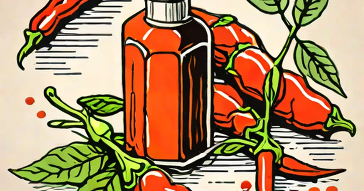
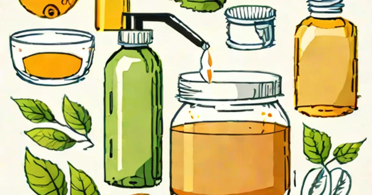
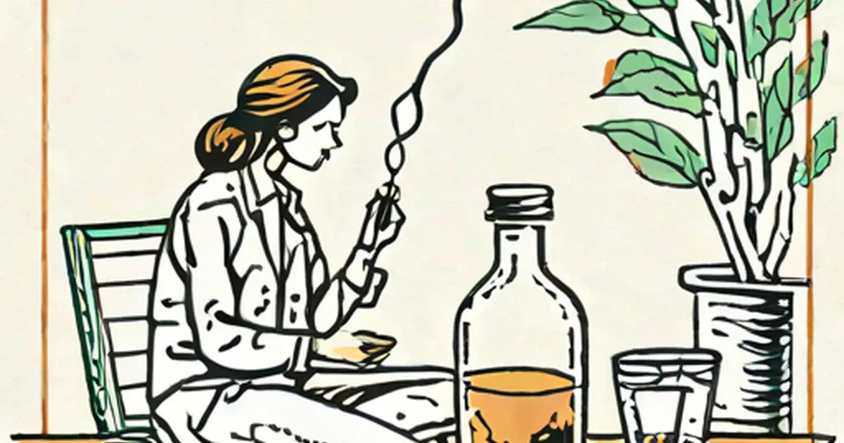
Does Apple Cider Vinegar Actually Help Acid Reflux? I Tried It
July 12, 2026
Articles in Health & Fitness.
Health & Fitness
July 12, 2026
Health & Fitness
July 12, 2026
Health & Fitness
July 12, 2026

Health & Fitness
July 3, 2026

Health & Fitness
July 3, 2026
Health & Fitness
July 3, 2026
Health & Fitness
July 3, 2026
Health & Fitness
July 3, 2026
Health & Fitness
July 3, 2026
Health & Fitness
July 3, 2026
Health & Fitness
July 3, 2026
Health & Fitness
July 3, 2026
Health & Fitness
July 3, 2026
Health & Fitness
July 3, 2026
Health & Fitness
July 3, 2026
Health & Fitness
July 3, 2026
Health & Fitness
July 3, 2026
Health & Fitness
July 3, 2026
Health & Fitness
July 3, 2026
Health & Fitness
July 3, 2026
Health & Fitness
July 3, 2026
Health & Fitness
July 3, 2026

Health & Fitness
July 3, 2026
Health & Fitness
July 3, 2026

Health & Fitness
July 3, 2026
Health & Fitness
July 3, 2026

Health & Fitness
July 3, 2026

Health & Fitness
July 3, 2026
Health & Fitness
July 3, 2026
Health & Fitness
July 3, 2026
Health & Fitness
July 3, 2026

Health & Fitness
July 3, 2026
Health & Fitness
July 3, 2026

Health & Fitness
July 3, 2026

Health & Fitness
July 3, 2026
Health & Fitness
July 3, 2026
Health & Fitness
July 3, 2026
Health & Fitness
July 3, 2026
Health & Fitness
July 3, 2026
Health & Fitness
July 3, 2026
Health & Fitness
July 3, 2026
Health & Fitness
July 3, 2026
Health & Fitness
July 3, 2026
Health & Fitness
July 3, 2026
Health & Fitness
July 3, 2026

Health & Fitness
July 3, 2026

Health & Fitness
July 3, 2026
Health & Fitness
July 3, 2026
Health & Fitness
July 3, 2026
Health & Fitness
July 3, 2026
Health & Fitness
July 3, 2026

Health & Fitness
July 3, 2026
Health & Fitness
July 3, 2026
Health & Fitness
July 3, 2026
Health & Fitness
July 3, 2026
Health & Fitness
July 3, 2026
Health & Fitness
July 3, 2026
Health & Fitness
July 3, 2026
Health & Fitness
July 3, 2026
Health & Fitness
July 3, 2026
Health & Fitness
July 3, 2026
Health & Fitness
July 3, 2026
Health & Fitness
July 3, 2026
Health & Fitness
July 3, 2026

Health & Fitness
July 3, 2026
Health & Fitness
July 3, 2026
Health & Fitness
July 3, 2026
Health & Fitness
July 3, 2026
Health & Fitness
July 3, 2026
Health & Fitness
July 3, 2026
Health & Fitness
July 3, 2026
Health & Fitness
July 3, 2026
Health & Fitness
July 3, 2026

Health & Fitness
July 3, 2026

Health & Fitness
July 3, 2026
Health & Fitness
July 3, 2026
Health & Fitness
July 3, 2026
Health & Fitness
July 3, 2026
Health & Fitness
July 3, 2026

Health & Fitness
July 3, 2026
Health & Fitness
July 3, 2026

Health & Fitness
July 3, 2026
Health & Fitness
July 3, 2026

Health & Fitness
July 3, 2026
Health & Fitness
July 3, 2026

Health & Fitness
July 3, 2026
Health & Fitness
July 3, 2026
Health & Fitness
July 3, 2026

Health & Fitness
July 3, 2026
Health & Fitness
July 3, 2026
Health & Fitness
July 3, 2026
Health & Fitness
July 3, 2026
Health & Fitness
July 3, 2026
Health & Fitness
July 3, 2026

Health & Fitness
July 3, 2026
Health & Fitness
July 3, 2026

Health & Fitness
July 3, 2026
Health & Fitness
July 3, 2026

Health & Fitness
July 3, 2026
Health & Fitness
July 3, 2026
Health & Fitness
July 3, 2026
Health & Fitness
July 3, 2026

Health & Fitness
July 3, 2026

Health & Fitness
July 3, 2026
Health & Fitness
July 3, 2026
Health & Fitness
July 3, 2026
Health & Fitness
July 3, 2026
Health & Fitness
July 3, 2026

Health & Fitness
July 3, 2026

Health & Fitness
July 3, 2026
Health & Fitness
July 3, 2026
Health & Fitness
July 3, 2026

Health & Fitness
July 3, 2026
Health & Fitness
July 3, 2026
Health & Fitness
July 3, 2026
Health & Fitness
July 3, 2026

Health & Fitness
July 3, 2026
Health & Fitness
July 3, 2026
Health & Fitness
July 3, 2026
Health & Fitness
July 3, 2026
Health & Fitness
July 3, 2026
Health & Fitness
July 3, 2026

Health & Fitness
July 3, 2026
Health & Fitness
July 3, 2026
Health & Fitness
July 3, 2026
Health & Fitness
July 3, 2026
Health & Fitness
July 3, 2026
Health & Fitness
July 3, 2026
Health & Fitness
July 3, 2026
Health & Fitness
July 3, 2026

Health & Fitness
July 3, 2026
Health & Fitness
July 3, 2026
Health & Fitness
July 3, 2026
Health & Fitness
July 3, 2026
Health & Fitness
July 3, 2026
Health & Fitness
July 3, 2026

Health & Fitness
July 3, 2026
Health & Fitness
July 3, 2026

Health & Fitness
July 3, 2026

Health & Fitness
July 3, 2026
Health & Fitness
July 3, 2026

Health & Fitness
July 3, 2026
Health & Fitness
July 3, 2026
Health & Fitness
July 3, 2026
Health & Fitness
July 3, 2026
Health & Fitness
July 3, 2026
Health & Fitness
July 3, 2026
Health & Fitness
July 3, 2026
Health & Fitness
July 3, 2026
Health & Fitness
July 3, 2026
Health & Fitness
July 3, 2026
Health & Fitness
July 3, 2026

Health & Fitness
July 3, 2026
Health & Fitness
July 3, 2026

Health & Fitness
July 3, 2026
Health & Fitness
July 3, 2026
Health & Fitness
July 3, 2026
Health & Fitness
July 3, 2026
Health & Fitness
July 3, 2026
Health & Fitness
July 3, 2026
Health & Fitness
July 3, 2026
Health & Fitness
July 3, 2026
Health & Fitness
July 3, 2026

Health & Fitness
July 3, 2026

Health & Fitness
July 3, 2026
Health & Fitness
July 3, 2026
Health & Fitness
July 3, 2026
Health & Fitness
July 3, 2026
Health & Fitness
July 3, 2026
Health & Fitness
July 3, 2026
Health & Fitness
July 3, 2026
Health & Fitness
July 3, 2026
Health & Fitness
July 3, 2026
Health & Fitness
July 3, 2026

Health & Fitness
July 3, 2026

Health & Fitness
July 3, 2026
Health & Fitness
July 3, 2026

Health & Fitness
July 3, 2026
Health & Fitness
July 3, 2026
Health & Fitness
July 3, 2026

Health & Fitness
July 3, 2026
Health & Fitness
July 3, 2026
Health & Fitness
July 3, 2026
Health & Fitness
July 3, 2026

Health & Fitness
July 3, 2026
Health & Fitness
July 3, 2026

Health & Fitness
July 3, 2026
Health & Fitness
July 3, 2026
Health & Fitness
July 3, 2026

Health & Fitness
July 3, 2026
Health & Fitness
July 3, 2026
Health & Fitness
July 3, 2026
Health & Fitness
July 3, 2026
Health & Fitness
July 3, 2026
Health & Fitness
July 3, 2026
Health & Fitness
July 3, 2026
Health & Fitness
July 3, 2026
Health & Fitness
July 3, 2026
Health & Fitness
July 3, 2026
Health & Fitness
July 3, 2026

Health & Fitness
July 3, 2026
Health & Fitness
July 3, 2026
Health & Fitness
July 3, 2026
Health & Fitness
July 3, 2026
Health & Fitness
July 3, 2026
Health & Fitness
July 3, 2026
Health & Fitness
July 3, 2026
Health & Fitness
July 3, 2026
Health & Fitness
July 3, 2026
Health & Fitness
July 3, 2026
Health & Fitness
July 3, 2026
Health & Fitness
July 3, 2026
Health & Fitness
July 3, 2026
Health & Fitness
July 3, 2026
Health & Fitness
July 3, 2026
Health & Fitness
July 3, 2026
Health & Fitness
July 3, 2026

Health & Fitness
July 3, 2026
Health & Fitness
July 3, 2026
Health & Fitness
July 3, 2026
Health & Fitness
July 3, 2026
Health & Fitness
July 3, 2026
Health & Fitness
July 3, 2026
Health & Fitness
July 3, 2026
Health & Fitness
July 3, 2026
Health & Fitness
July 3, 2026
Health & Fitness
July 3, 2026
Health & Fitness
July 3, 2026
Health & Fitness
July 3, 2026
Health & Fitness
July 3, 2026
Health & Fitness
July 3, 2026
Health & Fitness
July 3, 2026
Health & Fitness
July 3, 2026
Health & Fitness
July 3, 2026
Health & Fitness
July 3, 2026
Health & Fitness
July 3, 2026
Health & Fitness
July 3, 2026

Health & Fitness
July 3, 2026
Health & Fitness
July 3, 2026
Health & Fitness
July 3, 2026

Health & Fitness
July 3, 2026
Health & Fitness
July 3, 2026
Health & Fitness
July 3, 2026
Health & Fitness
July 3, 2026
Health & Fitness
July 3, 2026
Health & Fitness
July 3, 2026

Health & Fitness
July 3, 2026
Health & Fitness
July 3, 2026
Health & Fitness
July 3, 2026

Health & Fitness
July 3, 2026
Health & Fitness
July 3, 2026

Health & Fitness
July 3, 2026
Health & Fitness
July 3, 2026
Health & Fitness
July 3, 2026
Health & Fitness
July 3, 2026
Health & Fitness
July 3, 2026

Health & Fitness
July 3, 2026
Health & Fitness
July 3, 2026
Health & Fitness
July 3, 2026
Health & Fitness
July 3, 2026
Health & Fitness
July 3, 2026
Health & Fitness
July 3, 2026
Health & Fitness
July 3, 2026
Health & Fitness
July 3, 2026
Health & Fitness
July 3, 2026
Health & Fitness
July 3, 2026

Health & Fitness
July 3, 2026
Health & Fitness
July 3, 2026
Health & Fitness
July 3, 2026
Health & Fitness
July 3, 2026
Health & Fitness
July 3, 2026

Health & Fitness
July 3, 2026
Health & Fitness
July 3, 2026
Health & Fitness
July 3, 2026
Health & Fitness
July 3, 2026
Health & Fitness
July 3, 2026
Health & Fitness
July 3, 2026

Health & Fitness
July 3, 2026
Health & Fitness
July 3, 2026
Health & Fitness
July 3, 2026
Health & Fitness
July 3, 2026
Health & Fitness
July 3, 2026
Health & Fitness
July 3, 2026
Health & Fitness
July 3, 2026
Health & Fitness
July 3, 2026
Health & Fitness
July 3, 2026
Health & Fitness
July 3, 2026
Health & Fitness
July 3, 2026
Health & Fitness
July 3, 2026

Health & Fitness
July 3, 2026
Health & Fitness
July 3, 2026

Health & Fitness
July 3, 2026
Health & Fitness
July 3, 2026
Health & Fitness
July 3, 2026
Health & Fitness
July 3, 2026
Health & Fitness
July 3, 2026
Health & Fitness
July 3, 2026
Health & Fitness
July 3, 2026

Health & Fitness
July 3, 2026

Health & Fitness
July 3, 2026
Health & Fitness
July 3, 2026
Health & Fitness
July 3, 2026
Health & Fitness
July 3, 2026
Health & Fitness
July 3, 2026
Health & Fitness
July 3, 2026
Health & Fitness
July 3, 2026
Health & Fitness
July 3, 2026
Health & Fitness
July 3, 2026
Health & Fitness
July 3, 2026
Health & Fitness
July 3, 2026
Health & Fitness
July 3, 2026
Health & Fitness
July 3, 2026

Health & Fitness
July 3, 2026

Health & Fitness
July 3, 2026
Health & Fitness
July 3, 2026
Health & Fitness
July 3, 2026
Health & Fitness
July 3, 2026
Health & Fitness
July 3, 2026

Health & Fitness
July 3, 2026

Health & Fitness
July 3, 2026
Health & Fitness
July 3, 2026
Health & Fitness
July 3, 2026
Health & Fitness
July 3, 2026

Health & Fitness
July 3, 2026
Health & Fitness
July 3, 2026
Health & Fitness
July 3, 2026
Health & Fitness
July 3, 2026
Health & Fitness
July 3, 2026
Health & Fitness
July 3, 2026
Health & Fitness
July 3, 2026
Health & Fitness
July 3, 2026
Health & Fitness
July 3, 2026
Health & Fitness
July 3, 2026
Health & Fitness
July 3, 2026
Health & Fitness
July 3, 2026
Health & Fitness
July 3, 2026
Health & Fitness
July 3, 2026
Health & Fitness
July 3, 2026
Health & Fitness
July 3, 2026
Health & Fitness
July 3, 2026

Health & Fitness
July 3, 2026
Health & Fitness
July 3, 2026
Health & Fitness
July 3, 2026

Health & Fitness
July 3, 2026

Health & Fitness
July 3, 2026
Health & Fitness
July 3, 2026
Health & Fitness
July 3, 2026
Health & Fitness
July 3, 2026
Health & Fitness
July 3, 2026
Health & Fitness
July 3, 2026
Health & Fitness
July 3, 2026

Health & Fitness
July 3, 2026

Health & Fitness
July 3, 2026
Health & Fitness
July 3, 2026
Health & Fitness
July 3, 2026

Health & Fitness
July 3, 2026