Health & Fitness
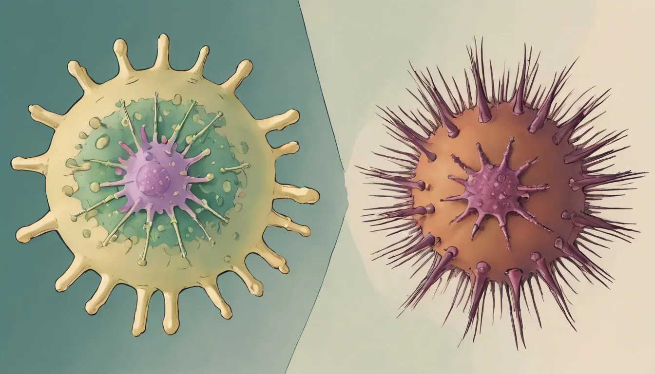
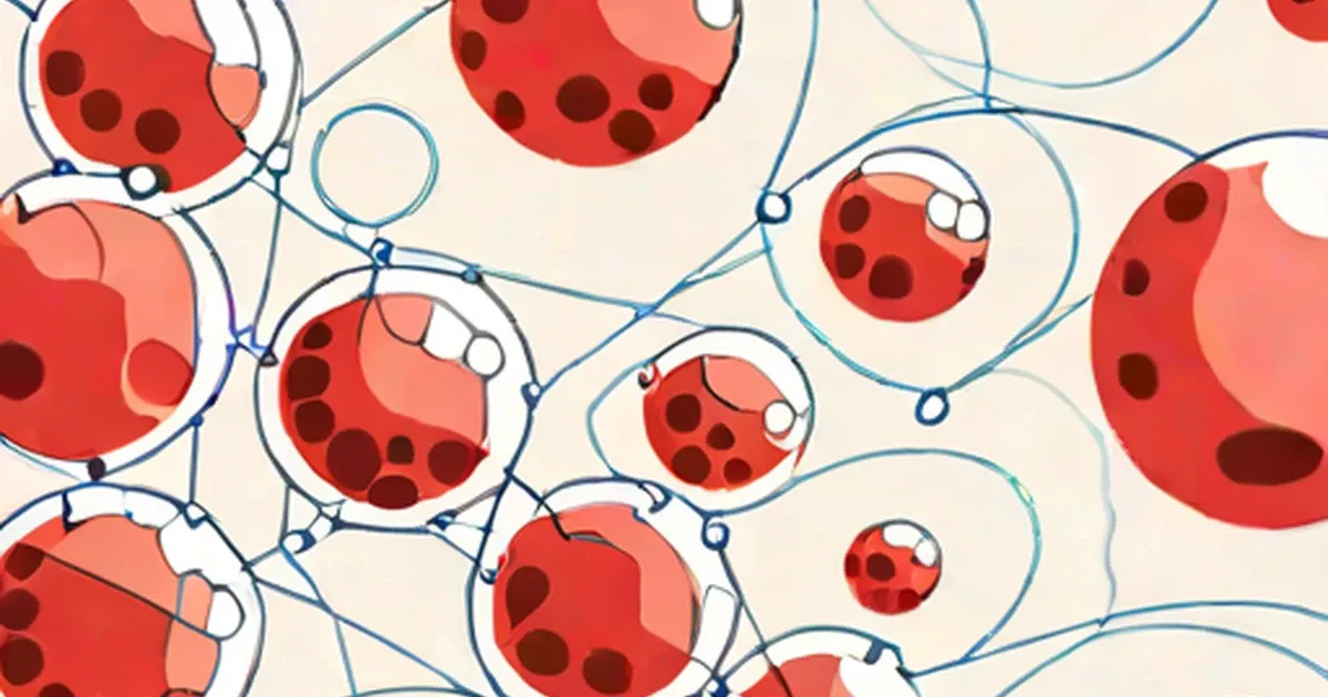
Innate vs. Adaptive Immunity: Your Body’s Two Defenses
July 18, 2026
Articles in Health & Fitness.
Health & Fitness
July 18, 2026
Health & Fitness
July 18, 2026
Health & Fitness
July 18, 2026
Health & Fitness
July 18, 2026
Health & Fitness
July 18, 2026

Health & Fitness
July 18, 2026
Health & Fitness
July 18, 2026
Health & Fitness
July 18, 2026
Health & Fitness
July 18, 2026
Health & Fitness
July 18, 2026
Health & Fitness
July 18, 2026
Health & Fitness
July 18, 2026
Health & Fitness
July 18, 2026
Health & Fitness
July 18, 2026
Health & Fitness
July 18, 2026
Health & Fitness
July 17, 2026
Health & Fitness
July 17, 2026
Health & Fitness
July 17, 2026
Health & Fitness
July 17, 2026
Health & Fitness
July 17, 2026
Health & Fitness
July 17, 2026
Health & Fitness
July 17, 2026

Health & Fitness
July 17, 2026

Health & Fitness
July 17, 2026

Health & Fitness
July 17, 2026

Health & Fitness
July 17, 2026

Health & Fitness
July 17, 2026

Health & Fitness
July 17, 2026
Health & Fitness
July 16, 2026
Health & Fitness
July 12, 2026
Health & Fitness
July 12, 2026
Health & Fitness
July 12, 2026

Health & Fitness
July 3, 2026

Health & Fitness
July 3, 2026
Health & Fitness
July 3, 2026
Health & Fitness
July 3, 2026
Health & Fitness
July 3, 2026
Health & Fitness
July 3, 2026
Health & Fitness
July 3, 2026

Health & Fitness
July 3, 2026
Health & Fitness
July 3, 2026
Health & Fitness
July 3, 2026
Health & Fitness
July 3, 2026
Health & Fitness
July 3, 2026
Health & Fitness
July 3, 2026
Health & Fitness
July 3, 2026
Health & Fitness
July 3, 2026
Health & Fitness
July 3, 2026
Health & Fitness
July 3, 2026
Health & Fitness
July 3, 2026
Health & Fitness
July 3, 2026

Health & Fitness
July 3, 2026
Health & Fitness
July 3, 2026

Health & Fitness
July 3, 2026
Health & Fitness
July 3, 2026

Health & Fitness
July 3, 2026

Health & Fitness
July 3, 2026
Health & Fitness
July 3, 2026
Health & Fitness
July 3, 2026

Health & Fitness
July 3, 2026

Health & Fitness
July 3, 2026
Health & Fitness
July 3, 2026

Health & Fitness
July 3, 2026

Health & Fitness
July 3, 2026
Health & Fitness
July 3, 2026
Health & Fitness
July 3, 2026
Health & Fitness
July 3, 2026
Health & Fitness
July 3, 2026
Health & Fitness
July 3, 2026
Health & Fitness
July 3, 2026
Health & Fitness
July 3, 2026
Health & Fitness
July 3, 2026

Health & Fitness
July 3, 2026
Health & Fitness
July 3, 2026

Health & Fitness
July 3, 2026

Health & Fitness
July 3, 2026
Health & Fitness
July 3, 2026
Health & Fitness
July 3, 2026
Health & Fitness
July 3, 2026
Health & Fitness
July 3, 2026

Health & Fitness
July 3, 2026
Health & Fitness
July 3, 2026
Health & Fitness
July 3, 2026
Health & Fitness
July 3, 2026
Health & Fitness
July 3, 2026
Health & Fitness
July 3, 2026
Health & Fitness
July 3, 2026
Health & Fitness
July 3, 2026
Health & Fitness
July 3, 2026
Health & Fitness
July 3, 2026
Health & Fitness
July 3, 2026
Health & Fitness
July 3, 2026
Health & Fitness
July 3, 2026

Health & Fitness
July 3, 2026
Health & Fitness
July 3, 2026
Health & Fitness
July 3, 2026
Health & Fitness
July 3, 2026
Health & Fitness
July 3, 2026
Health & Fitness
July 3, 2026
Health & Fitness
July 3, 2026
Health & Fitness
July 3, 2026
Health & Fitness
July 3, 2026

Health & Fitness
July 3, 2026

Health & Fitness
July 3, 2026
Health & Fitness
July 3, 2026
Health & Fitness
July 3, 2026
Health & Fitness
July 3, 2026
Health & Fitness
July 3, 2026

Health & Fitness
July 3, 2026
Health & Fitness
July 3, 2026

Health & Fitness
July 3, 2026
Health & Fitness
July 3, 2026

Health & Fitness
July 3, 2026
Health & Fitness
July 3, 2026

Health & Fitness
July 3, 2026
Health & Fitness
July 3, 2026
Health & Fitness
July 3, 2026

Health & Fitness
July 3, 2026
Health & Fitness
July 3, 2026
Health & Fitness
July 3, 2026
Health & Fitness
July 3, 2026
Health & Fitness
July 3, 2026
Health & Fitness
July 3, 2026

Health & Fitness
July 3, 2026
Health & Fitness
July 3, 2026

Health & Fitness
July 3, 2026
Health & Fitness
July 3, 2026

Health & Fitness
July 3, 2026
Health & Fitness
July 3, 2026
Health & Fitness
July 3, 2026
Health & Fitness
July 3, 2026

Health & Fitness
July 3, 2026

Health & Fitness
July 3, 2026
Health & Fitness
July 3, 2026
Health & Fitness
July 3, 2026
Health & Fitness
July 3, 2026
Health & Fitness
July 3, 2026

Health & Fitness
July 3, 2026

Health & Fitness
July 3, 2026
Health & Fitness
July 3, 2026
Health & Fitness
July 3, 2026

Health & Fitness
July 3, 2026
Health & Fitness
July 3, 2026
Health & Fitness
July 3, 2026

Health & Fitness
July 3, 2026

Health & Fitness
July 3, 2026
Health & Fitness
July 3, 2026
Health & Fitness
July 3, 2026
Health & Fitness
July 3, 2026
Health & Fitness
July 3, 2026
Health & Fitness
July 3, 2026

Health & Fitness
July 3, 2026
Health & Fitness
July 3, 2026
Health & Fitness
July 3, 2026
Health & Fitness
July 3, 2026
Health & Fitness
July 3, 2026
Health & Fitness
July 3, 2026
Health & Fitness
July 3, 2026
Health & Fitness
July 3, 2026

Health & Fitness
July 3, 2026
Health & Fitness
July 3, 2026
Health & Fitness
July 3, 2026
Health & Fitness
July 3, 2026
Health & Fitness
July 3, 2026
Health & Fitness
July 3, 2026

Health & Fitness
July 3, 2026
Health & Fitness
July 3, 2026

Health & Fitness
July 3, 2026

Health & Fitness
July 3, 2026
Health & Fitness
July 3, 2026

Health & Fitness
July 3, 2026
Health & Fitness
July 3, 2026
Health & Fitness
July 3, 2026
Health & Fitness
July 3, 2026
Health & Fitness
July 3, 2026
Health & Fitness
July 3, 2026
Health & Fitness
July 3, 2026

Health & Fitness
July 3, 2026
Health & Fitness
July 3, 2026
Health & Fitness
July 3, 2026
Health & Fitness
July 3, 2026

Health & Fitness
July 3, 2026
Health & Fitness
July 3, 2026

Health & Fitness
July 3, 2026
Health & Fitness
July 3, 2026
Health & Fitness
July 3, 2026
Health & Fitness
July 3, 2026
Health & Fitness
July 3, 2026
Health & Fitness
July 3, 2026
Health & Fitness
July 3, 2026
Health & Fitness
July 3, 2026
Health & Fitness
July 3, 2026

Health & Fitness
July 3, 2026

Health & Fitness
July 3, 2026
Health & Fitness
July 3, 2026
Health & Fitness
July 3, 2026
Health & Fitness
July 3, 2026
Health & Fitness
July 3, 2026
Health & Fitness
July 3, 2026
Health & Fitness
July 3, 2026
Health & Fitness
July 3, 2026
Health & Fitness
July 3, 2026
Health & Fitness
July 3, 2026

Health & Fitness
July 3, 2026

Health & Fitness
July 3, 2026
Health & Fitness
July 3, 2026

Health & Fitness
July 3, 2026
Health & Fitness
July 3, 2026
Health & Fitness
July 3, 2026

Health & Fitness
July 3, 2026

Health & Fitness
July 3, 2026
Health & Fitness
July 3, 2026
Health & Fitness
July 3, 2026

Health & Fitness
July 3, 2026
Health & Fitness
July 3, 2026

Health & Fitness
July 3, 2026
Health & Fitness
July 3, 2026
Health & Fitness
July 3, 2026

Health & Fitness
July 3, 2026
Health & Fitness
July 3, 2026
Health & Fitness
July 3, 2026
Health & Fitness
July 3, 2026

Health & Fitness
July 3, 2026
Health & Fitness
July 3, 2026
Health & Fitness
July 3, 2026
Health & Fitness
July 3, 2026
Health & Fitness
July 3, 2026
Health & Fitness
July 3, 2026
Health & Fitness
July 3, 2026

Health & Fitness
July 3, 2026
Health & Fitness
July 3, 2026
Health & Fitness
July 3, 2026
Health & Fitness
July 3, 2026
Health & Fitness
July 3, 2026
Health & Fitness
July 3, 2026
Health & Fitness
July 3, 2026
Health & Fitness
July 3, 2026

Health & Fitness
July 3, 2026
Health & Fitness
July 3, 2026
Health & Fitness
July 3, 2026
Health & Fitness
July 3, 2026
Health & Fitness
July 3, 2026
Health & Fitness
July 3, 2026
Health & Fitness
July 3, 2026
Health & Fitness
July 3, 2026
Health & Fitness
July 3, 2026

Health & Fitness
July 3, 2026
Health & Fitness
July 3, 2026
Health & Fitness
July 3, 2026
Health & Fitness
July 3, 2026
Health & Fitness
July 3, 2026
Health & Fitness
July 3, 2026
Health & Fitness
July 3, 2026
Health & Fitness
July 3, 2026
Health & Fitness
July 3, 2026
Health & Fitness
July 3, 2026
Health & Fitness
July 3, 2026
Health & Fitness
July 3, 2026
Health & Fitness
July 3, 2026
Health & Fitness
July 3, 2026
Health & Fitness
July 3, 2026
Health & Fitness
July 3, 2026
Health & Fitness
July 3, 2026
Health & Fitness
July 3, 2026
Health & Fitness
July 3, 2026
Health & Fitness
July 3, 2026

Health & Fitness
July 3, 2026
Health & Fitness
July 3, 2026
Health & Fitness
July 3, 2026

Health & Fitness
July 3, 2026
Health & Fitness
July 3, 2026
Health & Fitness
July 3, 2026
Health & Fitness
July 3, 2026
Health & Fitness
July 3, 2026
Health & Fitness
July 3, 2026

Health & Fitness
July 3, 2026
Health & Fitness
July 3, 2026

Health & Fitness
July 3, 2026

Health & Fitness
July 3, 2026
Health & Fitness
July 3, 2026

Health & Fitness
July 3, 2026
Health & Fitness
July 3, 2026
Health & Fitness
July 3, 2026
Health & Fitness
July 3, 2026
Health & Fitness
July 3, 2026

Health & Fitness
July 3, 2026
Health & Fitness
July 3, 2026
Health & Fitness
July 3, 2026
Health & Fitness
July 3, 2026
Health & Fitness
July 3, 2026
Health & Fitness
July 3, 2026
Health & Fitness
July 3, 2026

Health & Fitness
July 3, 2026
Health & Fitness
July 3, 2026
Health & Fitness
July 3, 2026

Health & Fitness
July 3, 2026
Health & Fitness
July 3, 2026
Health & Fitness
July 3, 2026
Health & Fitness
July 3, 2026
Health & Fitness
July 3, 2026

Health & Fitness
July 3, 2026
Health & Fitness
July 3, 2026
Health & Fitness
July 3, 2026
Health & Fitness
July 3, 2026
Health & Fitness
July 3, 2026

Health & Fitness
July 3, 2026

Health & Fitness
July 3, 2026
Health & Fitness
July 3, 2026
Health & Fitness
July 3, 2026
Health & Fitness
July 3, 2026
Health & Fitness
July 3, 2026
Health & Fitness
July 3, 2026
Health & Fitness
July 3, 2026

Health & Fitness
July 3, 2026
Health & Fitness
July 3, 2026
Health & Fitness
July 3, 2026
Health & Fitness
July 3, 2026

Health & Fitness
July 3, 2026
Health & Fitness
July 3, 2026

Health & Fitness
July 3, 2026
Health & Fitness
July 3, 2026
Health & Fitness
July 3, 2026
Health & Fitness
July 3, 2026

Health & Fitness
July 3, 2026
Health & Fitness
July 3, 2026
Health & Fitness
July 3, 2026

Health & Fitness
July 3, 2026

Health & Fitness
July 3, 2026
Health & Fitness
July 3, 2026
Health & Fitness
July 3, 2026
Health & Fitness
July 3, 2026
Health & Fitness
July 3, 2026
Health & Fitness
July 3, 2026
Health & Fitness
July 3, 2026
Health & Fitness
July 3, 2026
Health & Fitness
July 3, 2026
Health & Fitness
July 3, 2026
Health & Fitness
July 3, 2026
Health & Fitness
July 3, 2026
Health & Fitness
July 3, 2026

Health & Fitness
July 3, 2026

Health & Fitness
July 3, 2026
Health & Fitness
July 3, 2026
Health & Fitness
July 3, 2026
Health & Fitness
July 3, 2026
Health & Fitness
July 3, 2026

Health & Fitness
July 3, 2026

Health & Fitness
July 3, 2026
Health & Fitness
July 3, 2026
Health & Fitness
July 3, 2026
Health & Fitness
July 3, 2026

Health & Fitness
July 3, 2026
Health & Fitness
July 3, 2026
Health & Fitness
July 3, 2026
Health & Fitness
July 3, 2026
Health & Fitness
July 3, 2026
Health & Fitness
July 3, 2026
Health & Fitness
July 3, 2026
Health & Fitness
July 3, 2026
Health & Fitness
July 3, 2026
Health & Fitness
July 3, 2026
Health & Fitness
July 3, 2026
Health & Fitness
July 3, 2026
Health & Fitness
July 3, 2026
Health & Fitness
July 3, 2026
Health & Fitness
July 3, 2026
Health & Fitness
July 3, 2026
Health & Fitness
July 3, 2026

Health & Fitness
July 3, 2026
Health & Fitness
July 3, 2026
Health & Fitness
July 3, 2026

Health & Fitness
July 3, 2026

Health & Fitness
July 3, 2026
Health & Fitness
July 3, 2026
Health & Fitness
July 3, 2026
Health & Fitness
July 3, 2026
Health & Fitness
July 3, 2026
Health & Fitness
July 3, 2026
Health & Fitness
July 3, 2026

Health & Fitness
July 3, 2026

Health & Fitness
July 3, 2026
Health & Fitness
July 3, 2026
Health & Fitness
July 3, 2026

Health & Fitness
July 3, 2026