
Health & Fitness
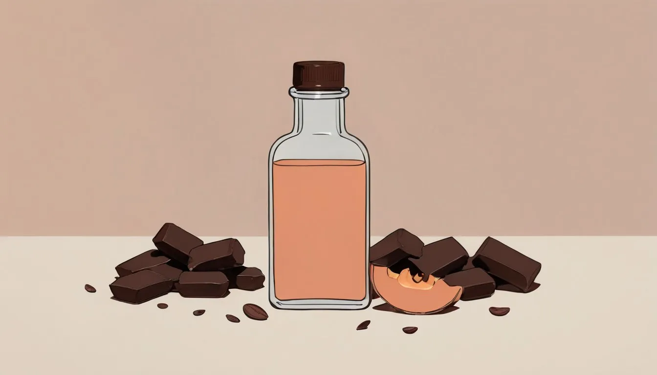
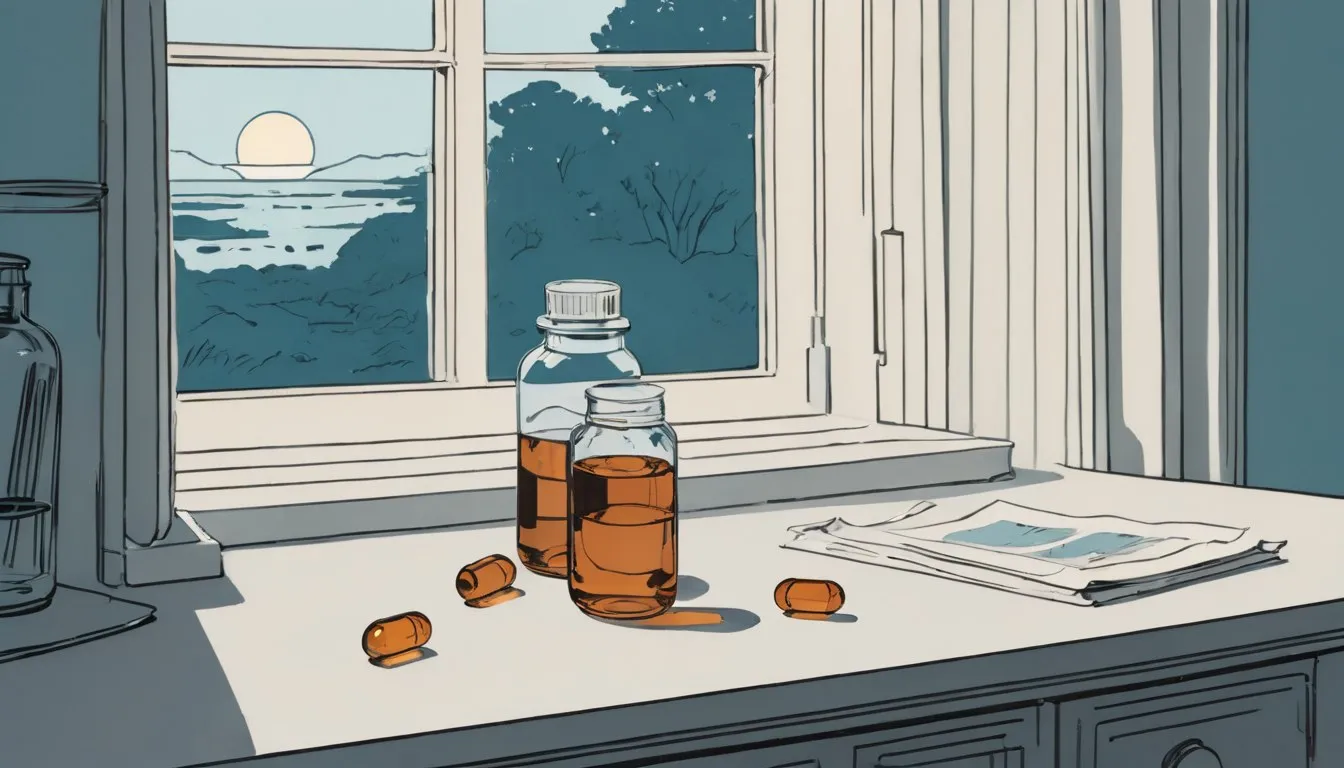
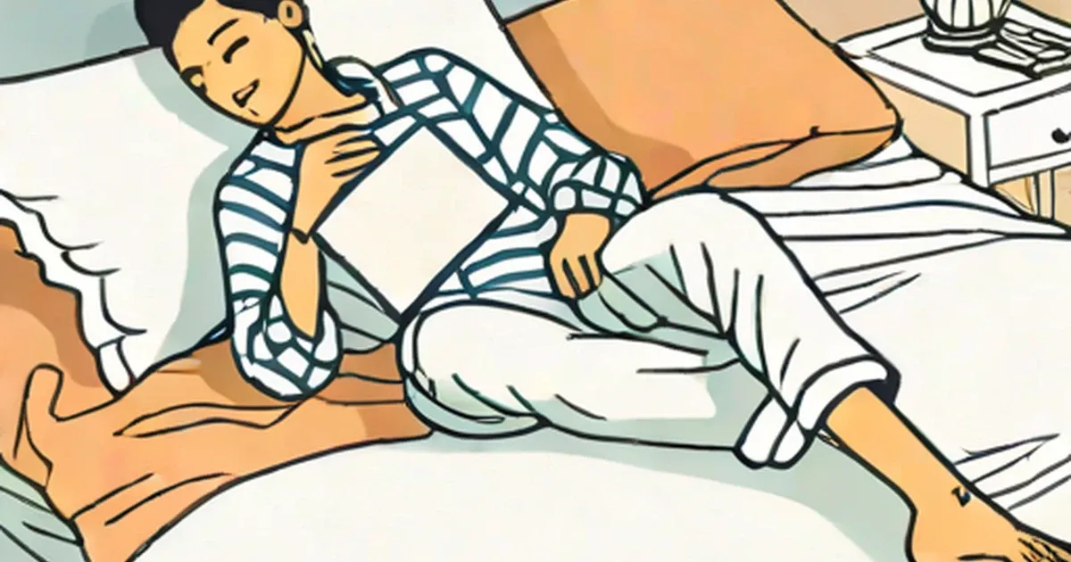
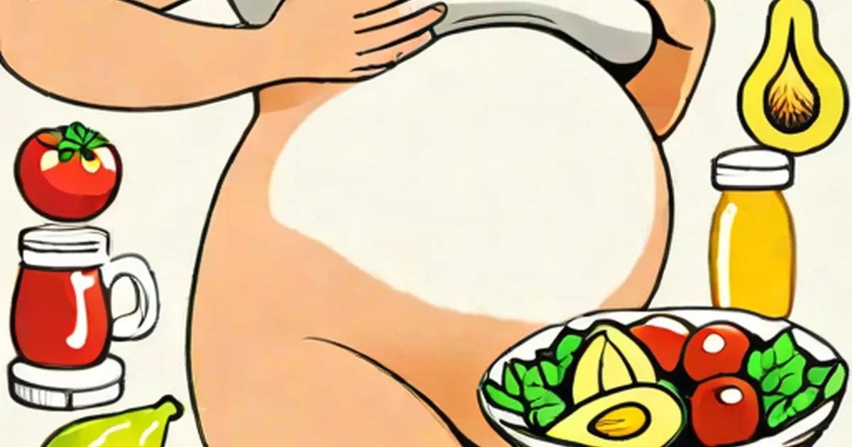
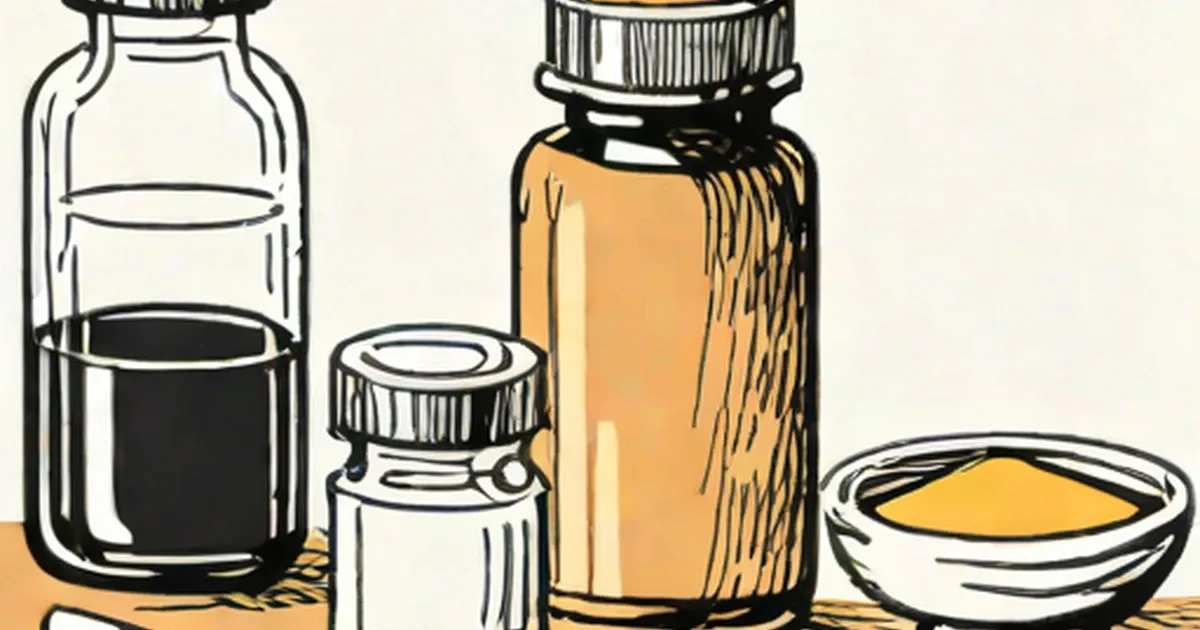
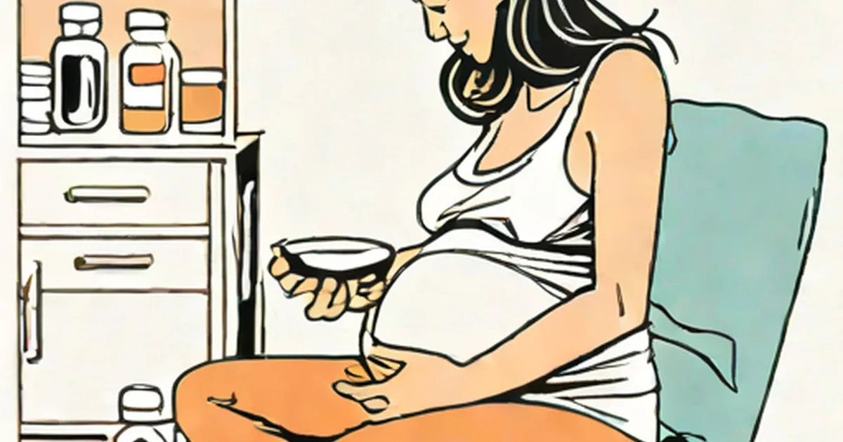
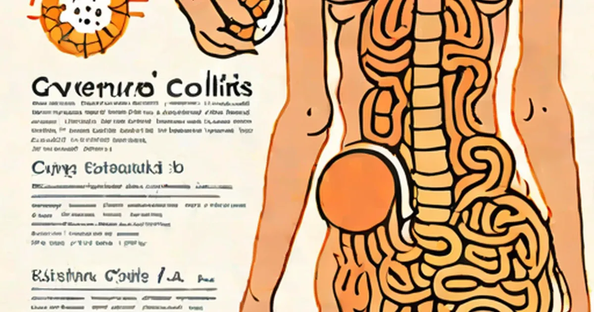
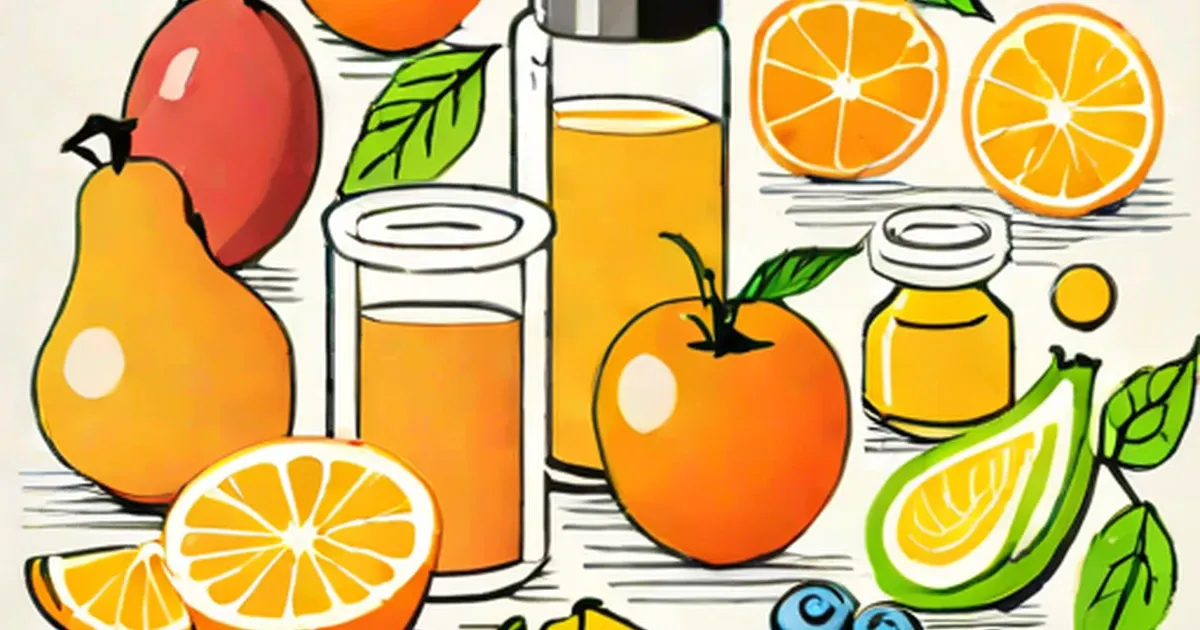
Is It Safe to Take Magnesium Supplements While Pregnant?
July 21, 2026
Health and fitness you can act on — nutrition, exercise, conditions and wellbeing, explained clearly and grounded in what the evidence supports.
Health & Fitness
July 21, 2026

Health & Fitness
July 21, 2026
Health & Fitness
July 21, 2026
Health & Fitness
July 21, 2026
Health & Fitness
July 21, 2026

Health & Fitness
July 21, 2026

Health & Fitness
July 21, 2026
Health & Fitness
July 21, 2026
Health & Fitness
July 21, 2026
Health & Fitness
July 21, 2026
Health & Fitness
July 21, 2026
Health & Fitness
July 21, 2026
Health & Fitness
July 21, 2026
Health & Fitness
July 21, 2026
Health & Fitness
July 21, 2026

Health & Fitness
July 21, 2026
Health & Fitness
July 20, 2026
Health & Fitness
July 20, 2026
Health & Fitness
July 20, 2026
Health & Fitness
July 20, 2026

Health & Fitness
July 20, 2026

Health & Fitness
July 20, 2026
Health & Fitness
July 20, 2026
Health & Fitness
July 20, 2026
Health & Fitness
July 20, 2026
Health & Fitness
July 20, 2026
Health & Fitness
July 18, 2026
Health & Fitness
July 18, 2026
Health & Fitness
July 18, 2026
Health & Fitness
July 18, 2026
Health & Fitness
July 18, 2026

Health & Fitness
July 18, 2026
Health & Fitness
July 18, 2026
Health & Fitness
July 18, 2026
Health & Fitness
July 18, 2026
Health & Fitness
July 18, 2026
Health & Fitness
July 18, 2026
Health & Fitness
July 18, 2026
Health & Fitness
July 18, 2026
Health & Fitness
July 18, 2026
Health & Fitness
July 18, 2026
Health & Fitness
July 17, 2026
Health & Fitness
July 17, 2026
Health & Fitness
July 17, 2026
Health & Fitness
July 17, 2026
Health & Fitness
July 17, 2026
Health & Fitness
July 17, 2026
Health & Fitness
July 17, 2026

Health & Fitness
July 17, 2026

Health & Fitness
July 17, 2026

Health & Fitness
July 17, 2026

Health & Fitness
July 17, 2026

Health & Fitness
July 17, 2026

Health & Fitness
July 17, 2026
Health & Fitness
July 16, 2026
Health & Fitness
July 12, 2026
Health & Fitness
July 12, 2026
Health & Fitness
July 12, 2026

Health & Fitness
July 3, 2026

Health & Fitness
July 3, 2026
Health & Fitness
July 3, 2026
Health & Fitness
July 3, 2026
Health & Fitness
July 3, 2026
Health & Fitness
July 3, 2026
Health & Fitness
July 3, 2026

Health & Fitness
July 3, 2026
Health & Fitness
July 3, 2026
Health & Fitness
July 3, 2026
Health & Fitness
July 3, 2026

Health & Fitness
July 3, 2026
Health & Fitness
July 3, 2026

Health & Fitness
July 3, 2026
Health & Fitness
July 3, 2026
Health & Fitness
July 3, 2026
Health & Fitness
July 3, 2026
Health & Fitness
July 3, 2026
Health & Fitness
July 3, 2026

Health & Fitness
July 3, 2026
Health & Fitness
July 3, 2026

Health & Fitness
July 3, 2026
Health & Fitness
July 3, 2026

Health & Fitness
July 3, 2026

Health & Fitness
July 3, 2026
Health & Fitness
July 3, 2026
Health & Fitness
July 3, 2026

Health & Fitness
July 3, 2026

Health & Fitness
July 3, 2026

Health & Fitness
July 3, 2026

Health & Fitness
July 3, 2026

Health & Fitness
July 3, 2026
Health & Fitness
July 3, 2026
Health & Fitness
July 3, 2026
Health & Fitness
July 3, 2026
Health & Fitness
July 3, 2026
Health & Fitness
July 3, 2026

Health & Fitness
July 3, 2026
Health & Fitness
July 3, 2026
Health & Fitness
July 3, 2026
Health & Fitness
July 3, 2026
Health & Fitness
July 3, 2026

Health & Fitness
July 3, 2026

Health & Fitness
July 3, 2026
Health & Fitness
July 3, 2026
Health & Fitness
July 3, 2026
Health & Fitness
July 3, 2026
Health & Fitness
July 3, 2026

Health & Fitness
July 3, 2026
Health & Fitness
July 3, 2026
Health & Fitness
July 3, 2026
Health & Fitness
July 3, 2026
Health & Fitness
July 3, 2026
Health & Fitness
July 3, 2026
Health & Fitness
July 3, 2026
Health & Fitness
July 3, 2026
Health & Fitness
July 3, 2026
Health & Fitness
July 3, 2026

Health & Fitness
July 3, 2026
Health & Fitness
July 3, 2026
Health & Fitness
July 3, 2026

Health & Fitness
July 3, 2026
Health & Fitness
July 3, 2026
Health & Fitness
July 3, 2026
Health & Fitness
July 3, 2026
Health & Fitness
July 3, 2026
Health & Fitness
July 3, 2026

Health & Fitness
July 3, 2026

Health & Fitness
July 3, 2026

Health & Fitness
July 3, 2026

Health & Fitness
July 3, 2026

Health & Fitness
July 3, 2026
Health & Fitness
July 3, 2026
Health & Fitness
July 3, 2026
Health & Fitness
July 3, 2026
Health & Fitness
July 3, 2026

Health & Fitness
July 3, 2026

Health & Fitness
July 3, 2026

Health & Fitness
July 3, 2026

Health & Fitness
July 3, 2026

Health & Fitness
July 3, 2026
Health & Fitness
July 3, 2026

Health & Fitness
July 3, 2026
Health & Fitness
July 3, 2026
Health & Fitness
July 3, 2026

Health & Fitness
July 3, 2026
Health & Fitness
July 3, 2026
Health & Fitness
July 3, 2026
Health & Fitness
July 3, 2026
Health & Fitness
July 3, 2026
Health & Fitness
July 3, 2026

Health & Fitness
July 3, 2026
Health & Fitness
July 3, 2026

Health & Fitness
July 3, 2026
Health & Fitness
July 3, 2026

Health & Fitness
July 3, 2026
Health & Fitness
July 3, 2026

Health & Fitness
July 3, 2026

Health & Fitness
July 3, 2026

Health & Fitness
July 3, 2026

Health & Fitness
July 3, 2026
Health & Fitness
July 3, 2026
Health & Fitness
July 3, 2026

Health & Fitness
July 3, 2026
Health & Fitness
July 3, 2026

Health & Fitness
July 3, 2026

Health & Fitness
July 3, 2026
Health & Fitness
July 3, 2026

Health & Fitness
July 3, 2026

Health & Fitness
July 3, 2026
Health & Fitness
July 3, 2026
Health & Fitness
July 3, 2026

Health & Fitness
July 3, 2026

Health & Fitness
July 3, 2026
Health & Fitness
July 3, 2026
Health & Fitness
July 3, 2026
Health & Fitness
July 3, 2026
Health & Fitness
July 3, 2026
Health & Fitness
July 3, 2026

Health & Fitness
July 3, 2026
Health & Fitness
July 3, 2026
Health & Fitness
July 3, 2026
Health & Fitness
July 3, 2026
Health & Fitness
July 3, 2026
Health & Fitness
July 3, 2026
Health & Fitness
July 3, 2026
Health & Fitness
July 3, 2026

Health & Fitness
July 3, 2026
Health & Fitness
July 3, 2026

Health & Fitness
July 3, 2026
Health & Fitness
July 3, 2026

Health & Fitness
July 3, 2026
Health & Fitness
July 3, 2026

Health & Fitness
July 3, 2026
Health & Fitness
July 3, 2026

Health & Fitness
July 3, 2026

Health & Fitness
July 3, 2026
Health & Fitness
July 3, 2026

Health & Fitness
July 3, 2026
Health & Fitness
July 3, 2026
Health & Fitness
July 3, 2026
Health & Fitness
July 3, 2026
Health & Fitness
July 3, 2026

Health & Fitness
July 3, 2026
Health & Fitness
July 3, 2026

Health & Fitness
July 3, 2026

Health & Fitness
July 3, 2026
Health & Fitness
July 3, 2026

Health & Fitness
July 3, 2026

Health & Fitness
July 3, 2026
Health & Fitness
July 3, 2026

Health & Fitness
July 3, 2026

Health & Fitness
July 3, 2026
Health & Fitness
July 3, 2026
Health & Fitness
July 3, 2026
Health & Fitness
July 3, 2026

Health & Fitness
July 3, 2026

Health & Fitness
July 3, 2026

Health & Fitness
July 3, 2026
Health & Fitness
July 3, 2026

Health & Fitness
July 3, 2026

Health & Fitness
July 3, 2026
Health & Fitness
July 3, 2026
Health & Fitness
July 3, 2026
Health & Fitness
July 3, 2026
Health & Fitness
July 3, 2026
Health & Fitness
July 3, 2026
Health & Fitness
July 3, 2026
Health & Fitness
July 3, 2026
Health & Fitness
July 3, 2026
Health & Fitness
July 3, 2026

Health & Fitness
July 3, 2026

Health & Fitness
July 3, 2026
Health & Fitness
July 3, 2026

Health & Fitness
July 3, 2026
Health & Fitness
July 3, 2026
Health & Fitness
July 3, 2026

Health & Fitness
July 3, 2026

Health & Fitness
July 3, 2026
Health & Fitness
July 3, 2026
Health & Fitness
July 3, 2026

Health & Fitness
July 3, 2026

Health & Fitness
July 3, 2026

Health & Fitness
July 3, 2026
Health & Fitness
July 3, 2026

Health & Fitness
July 3, 2026

Health & Fitness
July 3, 2026
Health & Fitness
July 3, 2026
Health & Fitness
July 3, 2026
Health & Fitness
July 3, 2026

Health & Fitness
July 3, 2026

Health & Fitness
July 3, 2026
Health & Fitness
July 3, 2026
Health & Fitness
July 3, 2026
Health & Fitness
July 3, 2026
Health & Fitness
July 3, 2026
Health & Fitness
July 3, 2026

Health & Fitness
July 3, 2026
Health & Fitness
July 3, 2026
Health & Fitness
July 3, 2026
Health & Fitness
July 3, 2026
Health & Fitness
July 3, 2026
Health & Fitness
July 3, 2026
Health & Fitness
July 3, 2026
Health & Fitness
July 3, 2026

Health & Fitness
July 3, 2026
Health & Fitness
July 3, 2026
Health & Fitness
July 3, 2026
Health & Fitness
July 3, 2026
Health & Fitness
July 3, 2026
Health & Fitness
July 3, 2026
Health & Fitness
July 3, 2026
Health & Fitness
July 3, 2026
Health & Fitness
July 3, 2026

Health & Fitness
July 3, 2026
Health & Fitness
July 3, 2026
Health & Fitness
July 3, 2026
Health & Fitness
July 3, 2026
Health & Fitness
July 3, 2026
Health & Fitness
July 3, 2026
Health & Fitness
July 3, 2026
Health & Fitness
July 3, 2026
Health & Fitness
July 3, 2026
Health & Fitness
July 3, 2026
Health & Fitness
July 3, 2026
Health & Fitness
July 3, 2026
Health & Fitness
July 3, 2026
Health & Fitness
July 3, 2026

Health & Fitness
July 3, 2026
Health & Fitness
July 3, 2026
Health & Fitness
July 3, 2026
Health & Fitness
July 3, 2026
Health & Fitness
July 3, 2026
Health & Fitness
July 3, 2026

Health & Fitness
July 3, 2026
Health & Fitness
July 3, 2026
Health & Fitness
July 3, 2026

Health & Fitness
July 3, 2026
Health & Fitness
July 3, 2026
Health & Fitness
July 3, 2026
Health & Fitness
July 3, 2026
Health & Fitness
July 3, 2026

Health & Fitness
July 3, 2026

Health & Fitness
July 3, 2026

Health & Fitness
July 3, 2026

Health & Fitness
July 3, 2026

Health & Fitness
July 3, 2026
Health & Fitness
July 3, 2026

Health & Fitness
July 3, 2026
Health & Fitness
July 3, 2026
Health & Fitness
July 3, 2026
Health & Fitness
July 3, 2026
Health & Fitness
July 3, 2026

Health & Fitness
July 3, 2026
Health & Fitness
July 3, 2026
Health & Fitness
July 3, 2026
Health & Fitness
July 3, 2026
Health & Fitness
July 3, 2026
Health & Fitness
July 3, 2026
Health & Fitness
July 3, 2026

Health & Fitness
July 3, 2026
Health & Fitness
July 3, 2026
Health & Fitness
July 3, 2026

Health & Fitness
July 3, 2026
Health & Fitness
July 3, 2026
Health & Fitness
July 3, 2026
Health & Fitness
July 3, 2026
Health & Fitness
July 3, 2026

Health & Fitness
July 3, 2026
Health & Fitness
July 3, 2026
Health & Fitness
July 3, 2026
Health & Fitness
July 3, 2026
Health & Fitness
July 3, 2026

Health & Fitness
July 3, 2026

Health & Fitness
July 3, 2026
Health & Fitness
July 3, 2026

Health & Fitness
July 3, 2026
Health & Fitness
July 3, 2026

Health & Fitness
July 3, 2026

Health & Fitness
July 3, 2026

Health & Fitness
July 3, 2026

Health & Fitness
July 3, 2026
Health & Fitness
July 3, 2026
Health & Fitness
July 3, 2026

Health & Fitness
July 3, 2026

Health & Fitness
July 3, 2026

Health & Fitness
July 3, 2026

Health & Fitness
July 3, 2026
Health & Fitness
July 3, 2026
Health & Fitness
July 3, 2026
Health & Fitness
July 3, 2026

Health & Fitness
July 3, 2026
Health & Fitness
July 3, 2026
Health & Fitness
July 3, 2026

Health & Fitness
July 3, 2026

Health & Fitness
July 3, 2026
Health & Fitness
July 3, 2026
Health & Fitness
July 3, 2026
Health & Fitness
July 3, 2026
Health & Fitness
July 3, 2026
Health & Fitness
July 3, 2026
Health & Fitness
July 3, 2026

Health & Fitness
July 3, 2026

Health & Fitness
July 3, 2026
Health & Fitness
July 3, 2026
Health & Fitness
July 3, 2026
Health & Fitness
July 3, 2026
Health & Fitness
July 3, 2026

Health & Fitness
July 3, 2026

Health & Fitness
July 3, 2026

Health & Fitness
July 3, 2026
Health & Fitness
July 3, 2026
Health & Fitness
July 3, 2026
Health & Fitness
July 3, 2026

Health & Fitness
July 3, 2026

Health & Fitness
July 3, 2026
Health & Fitness
July 3, 2026
Health & Fitness
July 3, 2026
Health & Fitness
July 3, 2026

Health & Fitness
July 3, 2026
Health & Fitness
July 3, 2026
Health & Fitness
July 3, 2026
Health & Fitness
July 3, 2026
Health & Fitness
July 3, 2026
Health & Fitness
July 3, 2026
Health & Fitness
July 3, 2026
Health & Fitness
July 3, 2026
Health & Fitness
July 3, 2026
Health & Fitness
July 3, 2026
Health & Fitness
July 3, 2026
Health & Fitness
July 3, 2026
Health & Fitness
July 3, 2026
Health & Fitness
July 3, 2026

Health & Fitness
July 3, 2026
Health & Fitness
July 3, 2026
Health & Fitness
July 3, 2026

Health & Fitness
July 3, 2026
Health & Fitness
July 3, 2026
Health & Fitness
July 3, 2026

Health & Fitness
July 3, 2026

Health & Fitness
July 3, 2026
Health & Fitness
July 3, 2026
Health & Fitness
July 3, 2026
Health & Fitness
July 3, 2026
Health & Fitness
July 3, 2026

Health & Fitness
July 3, 2026
Health & Fitness
July 3, 2026

Health & Fitness
July 3, 2026

Health & Fitness
July 3, 2026
Health & Fitness
July 3, 2026
Health & Fitness
July 3, 2026

Health & Fitness
July 3, 2026